This is an automatically translated article.
The article is professionally consulted by Master, Doctor Le Hong Chien - Doctor of Radiology - Intervention - Department of Diagnostic Imaging and Nuclear Medicine - Vinmec Times City International General Hospital.Digital mesenteric angiography with background erasure is an imaging technique with an iodinated contrast medium to visualize the superior or inferior mesenteric vasculature. These are the arteries that supply blood to the small intestine, colon, and rectum.
1. When is the digitized mesenteric artery angiography indicated?
Digital angiography of the mesenteric arteries is indicated when:● When the doctor needs to assess the blood supply of the mesenteric artery
● Suspected mesenteric vascular disease: malformation, stenosis, embolism..
● Patient with gastrointestinal bleeding suspected of vascular malformation
● Patient with bleeding gastrointestinal tumor
● Need assessment of portal vein
● Perform angiography to serve for Interventional radiography
In some cases, the doctor needs to evaluate the patient's health status to decide whether to have digitalized mesenteric angiography, such as: The patient has a clotting disorder, kidney failure, money History of obvious allergy to iodinated contrast agents, pregnant women.
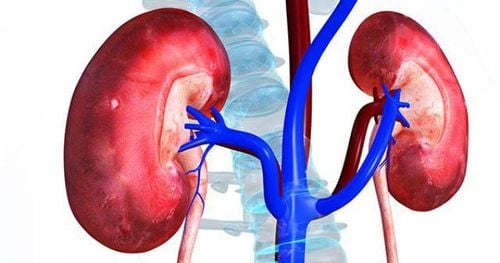
Bệnh nhân bị suy thận cần đánh giá tình trạng sức khoẻ trước khi quyết định có chụp động mạch mạc treo số hóa xóa nền không
2. Digital background erasing mesenteric angiography
To perform digital mesenteric angiography to erase the background, one specialist doctor, one nurse, and one electro-optical technician are required.An anesthesiologist is needed if the patient is uncooperative or the child is under 5 years of age.
2.1 Means of digitalized mesenteric angiography with background erasure
● Digital erasure angiography (DSA) machine● Specialized electric pump
Angiography kit, contrast agent
● Film, film printer, image storage system
● Lead jacket, impurities apron, shielding X-rays.
2.2 Patient
● The patient was thoroughly explained about the procedure to coordinate with the doctor in the process.● Fasting, drinking before 6 hours. You can drink water but not more than 50ml of water.
● At the intervention room: the patient lies on his back, installing a monitor to monitor breathing, pulse, blood pressure, electrocardiogram, SpO2. Disinfect the skin then spread a sterile intervention kit.

Máy chụp mạch số hóa xóa nền (DSA)
2.3 Perform digitized mesenteric angiography to erase the background
Place the patient supine on the imaging table, place an intravenous line (usually 0.9 % isotonic saline serum). Usually local anesthetic.2.4 Select the technique of use and the route of catheterization
● Arterial access using the Seldinger technique. Usually most of the time is from the femoral artery, unless this is not possible, other entrances from the axillary, brachial, or radial arteries are used.● Disinfection and local anesthetic.
Needle puncture and then insert the catheter
● Selective angiography of the superior mesenteric artery: Insert the 5F catheter into the abdominal aorta at the level of L1 -2, rotate the tip of the catheter anteriorly to hook into the choroidal artery. Mesenteric colon and then proceed to pump the drug at a rate of 4 -5ml/s, volume 12-16 ml, pump under 500PSI pressure.
● Super-selective microcatheter can be inserted into each branch of the mesenteric artery through the 5F catheter and then injected at a rate of 2ml/s, volume 6ml, pressure 250-300PSI.
● Lower mesenteric angiogram: Insert the 5F catheter into the abdominal aorta at the level of L3 - 4, rotate the tip of the catheter anteriorly slightly to the left to hook into the lower mesentery, and then proceed to pump the drug at high speed. 3ml/s, volume 6-9ml.
● Super-selective microcatheter can be inserted into each branch of the mesenteric artery through the 5F catheter and then injected at a rate of 2ml/s, volume 6ml, pressure 250-300PSI.
● The series focuses on the anterior - posterior direction of the mesenteric artery, taking the arterial, parenchymal and venous phases.
After satisfactory imaging, withdraw the catheter and tube into the lumen and then apply manual pressure directly on the needle puncture site for about 15 minutes to stop bleeding, then apply pressure for 6-8 hours or use an instrument to close the lumen .
The above is considered important information about the digital mesenteric angiography technique to erase the background. Customers can refer to the process to understand the steps.
Currently, Vinmec International General Hospital is a high-quality medical facility in Vietnam with a team of qualified, well-trained, domestic and foreign doctors and experienced doctors. . A system of modern and advanced medical equipment, possessing many of the best machines in the world, helps to detect many difficult and dangerous diseases in a short time. Thanks to good medical conditions, the results of diagnosis and treatment are always guaranteed to be the best for patients.
Master. Dr. Le Hong Chien has many years of experience working in the field of diagnostic imaging and interventional radiology (endovascular intervention and extravascular intervention). Before being a Doctor of Diagnostic Imaging - Intervention at Vinmec Times City International Hospital, Dr. Chien was a Doctor at the Department of Diagnostic Imaging, Hospital 19.8 - Ministry of Public Security and Hong Ngoc General Hospital. .
Any questions that need to be answered by a specialist doctor as well as customers wishing to be examined and treated at Vinmec International General Hospital, you can contact Vinmec Health System nationwide or register online HERE.
SEE MORE
Are mesenteric tumors dangerous? What is mesenteric lymphadenitis? Diagnosis and treatment of mesenteric lymphadenitis