
This is an automatically translated article.
The article was professionally consulted with resident doctor Nguyen Quynh Giang - Department of Diagnostic Imaging and Nuclear Medicine - Vinmec Times City International General Hospital.Currently, gastrointestinal disease is one of the common diseases. The introduction of the method of surveying the digestive tract plays a huge role in the diagnosis and treatment of digestive diseases, in which the gastrointestinal ultrasound technique is a widely used diagnostic tool. most today.
1. Examination of the digestive tract
Gastrointestinal diseases are a group of diseases that are encountered a lot in patients of all ages, genders and in any country. Therefore, the research on pathology, diagnostic means as well as effective treatment methods is always improved and enhanced every day with the desire to bring the best health to patients. In order to be able to survey the digestive tract and timely detect diseases appearing in these organs, a number of techniques and paraclinical methods have been born, contributing a great deal to doctors in the treatment of diseases. diagnose and find out the causes of the physical symptoms of the gastrointestinal tract upon clinical examination, with some typical methods such as gastrointestinal ultrasound, gastrointestinal endoscopy, abdominal computed tomography, Magnetic resonance imaging... In which, gastrointestinal ultrasound is the most popular method because it is easy to perform, affordable, fast and brings high efficiency in diagnosis and treatment monitoring.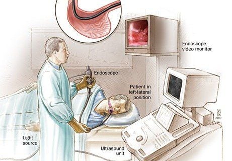
Nội soi đường tiêu hóa
2. Gastrointestinal ultrasound
Unlike other diagnostic methods of gastrointestinal diseases such as endoscopy or computed tomography, gastrointestinal ultrasound can give cross-sectional imaging results of the gastrointestinal tract wall and surrounding tissues. Some common pathologies that gastrointestinal ultrasound can detect are cases of gastrointestinal wall thickening, gastrointestinal wall tumor with or without ulceration, dilatation and collection of fluid around the intestinal loop or a number of soft tissues of the mesentery, great omentum:Cases of gastrointestinal wall thickening: Ultrasound can detect focal, diffuse, symmetric or asymmetrical gastrointestinal wall thickening images... with Images such as the pseudorenal sign, a target image characterized by an inferior echo ring on the outside, with a central echo, indicate that the bowel is narrowing or that an ulcer is present at the site. this. These results of thickening of the gastrointestinal tract wall can be directed to a number of pathological causes such as enteritis, colitis, intestinal cell hyperplasia, edema in pseudomembranous colitis, etc.
Case of gastrointestinal wall tumor:

Nếu khối u là do cơ trơn sẽ cho thấy những đặc điểm của hoại tử, tế bào thường đặc và bị nang hóa
Case of dilated and stagnant bowel loops: Dilated and stagnant bowel loops are the typical picture in intestinal obstruction or gastrointestinal obstruction. When intestinal obstruction occurs, there will be a lot of gas in the intestinal lumen, causing artifacts on ultrasound and it is difficult to detect these cases by gastrointestinal ultrasound. Therefore, it is necessary to remove gas as well as stagnant dilated bowel loops so that ultrasound can be performed most easily, helping to find intestinal obstruction and paralytic ileus in the patient as soon as possible.
Cases of soft tissue tumors in the mesentery and great omentum:
Đối với bệnh lý Crohn có u mô mềm xung quanh cần chụp cắt lớp vi tính để chẩn đoán
3. Conclusion
In order to diagnose as well as monitor the effectiveness of treatment of gastrointestinal diseases, it is indispensable to receive support from the methods of surveying the digestive tract, in which the easiest to perform is the extremely advanced gastrointestinal ultrasound technique. Fast, convenient and economical but still extremely effective.Vinmec International General Hospital applies gastrointestinal endoscopy, helping to accurately diagnose patients' digestive diseases, helping to provide the right treatment regimen, timely intervention, and limiting complications.
Vinmec is equipped with synchronously the most modern flexible endoscope system of Olympus - Japan, a prestigious manufacturer of the world's leading quality. The foot pedal irrigation system helps the doctor to take the initiative in irrigating the patient's gastrointestinal tract.
Please dial HOTLINE for more information or register for an appointment HERE. Download MyVinmec app to make appointments faster and to manage your bookings easily.