This is an automatically translated article.
The article is professionally consulted by Master, Doctor Nguyen Viet Thu - Doctor of Radiology and Nuclear Medicine - Department of Diagnostic Imaging and Nuclear Medicine - Vinmec Times City International General Hospital. Dr. Nguyen Viet Thu has more than 20 years of experience working in the field of diagnostic imaging, former Secretary of the Hanoi Department of Radiology, directing the lower level in the field of Diagnostic Imaging.Ultrasound is considered as one of the effective diagnostic tools in the evaluation and morphology of lymphoma. In addition, ultrasound plays an important role in monitoring the growth of lymph nodes.
1. What is a lymph node?
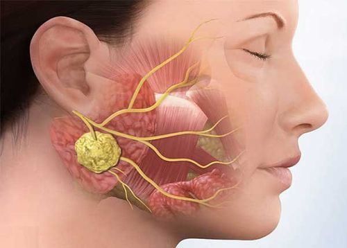
The human body has about 500 - 600 lymph nodes distributed throughout the body, they stand in groups and receive lymph from each body region.We can only feel or touch a few. Places where you can feel the lymph nodes are:
Lymph nodes in the angle of the jaw Lymph nodes behind the ear Lymph nodes in the axillary fossa Lymph nodes in the neck Inguinal lymph nodes Lymph nodes in the supraclavicular fos . The size of the lymph node usually varies, from as small as a pinhead to as large as a pea, which is part of the body's immune system.
Trắc nghiệm: Thử hiểu biết của bạn về hạch bạch huyết
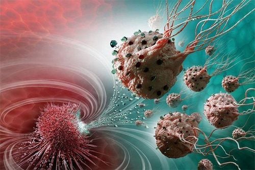
Hạch bạch huyết có vai trò quan trọng trong hệ miễn dịch của cơ thể. Bài trắc nghiệm dưới đây giúp bạn hiểu phần nào vai trò và chức năng của hạch bạch huyết.
Bài dịch từ: webmd.com
2. What is lymphoma?
Lymphoma is a condition in which the lymph nodes in the body are enlarged or swollen.
Bệnh u hạch ở trẻ
Although lymph node tumors are quite common, this does not mean that the disease will not cause any serious problems. Therefore, you must regularly go to the doctor for early and best diagnosis and treatment.
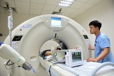
3. Ultrasound diagnosis of lymph node tumors by ultrasound
Ultrasound is a noninvasive and highly suggestive examination that is evaluable and useful for the diagnosis of lymph node diseases.Ultrasound can not only confirm or rule out the presence of a mass, but in many cases can also suggest the nature of the disease based on findings on ultrasound. Color Doppler ultrasound contributes significantly to increasing the reliability of the assessment of lymph node pathology.
3.1 Lymph node location The retroperitoneal lymph node is the most common in Lymphoma. Para-aortic lymphadenopathy is seen in 25% of Hodgkin's patients and 50% of non-Hodgkin's patients. 3.2 Shape May present with the phenotypes
Individual nodules oval, elongated, flattened or round. Gather in groups. The lymph nodes develop around a vascular axis consisting of arteries and veins. Mantle sign: The lymph nodes form a "jacket" that surrounds the great blood vessels. Floating aorta sign: Several retroperitoneal tumors or lymph nodes grow and spread along the retroperitoneal spaces, interlace between structures, and compress the abdominal aorta. “sandwich sign”: Blood vessels are sandwiched between clusters of lymphoma. 3.3 Resonance Fluid lymph nodes with empty echo and posterior echogenicity are often due to necrosis after treatment such as radiation therapy. Central gangrene or cystic fibrosis is usually caused by inflammation or tuberculosis. Mixed echogenic lymph nodes suggest lymph node metastasis. Calcification in the lymph nodes is uncommon and can be seen in lymph node tuberculosis. 3.4 Dimensions There is compression of surrounding structures. Perfusion (via color Doppler): Peripheral, central, or mixed distribution. Other signs: The effect of displacement or compression such as lymph nodes in the head of the pancreas, the hilum of the liver can compress the common bile duct. Lymph nodes around the inferior vena cava compress the blood vessels or large lymph nodes compress nearby organs. 3.5 Evaluation of benign - malignant nodes Benign nodes are detected usually with short axis diameter / long axis diameter < 0.5, corresponding to oval shape. Malignant nodes usually have this index > 0.5, which corresponds to a more rounded shape.
4. Diagnosis of Lymphoma Cause
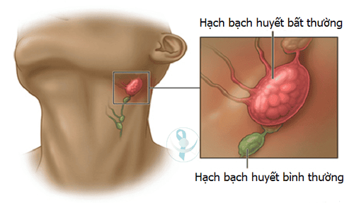
U hạch bạch huyết ở cổ
Ask for medical history. Nodal nature: Pain? Fast or slow development? Amount? Lymph node drainage. How to diagnose benign or malignant lymph nodes? After that, do other diagnostic methods such as CT scan, MRI (identify lymph nodes and evaluate cancer stage,...)? Select fine needle aspiration cytology (FNAC) or lymph node biopsy to diagnose the nodal nature.
In fact, the nature of lymph nodes can vary depending on the disease, making it difficult to distinguish between benign and malignant nodes. Therefore, as soon as there are symptoms of abnormally floating lymph nodes for no apparent reason, it is best for the patient to go to a reputable and professional medical facility for an accurate diagnosis. Depending on the location of the lymph nodes and the clinical examination results, the doctor may order tests such as ultrasound of the respective areas, CT scan, MRI scan, lymph node biopsy for cytology...
Please dial HOTLINE for more information or register for an appointment HERE. Download MyVinmec app to make appointments faster and to manage your bookings easily.