This is an automatically translated article.
The article was professionally consulted by Doctor Phan Dinh Thuy Tien - General Internal Medicine - Department of Examination & Internal Medicine - Vinmec Nha Trang International General HospitalAcute pulmonary embolism is an acute pulmonary embolism caused by a blood clot traveling from the deep veins of the legs to the lungs, leading to a potentially life-threatening pulmonary embolism. However, prompt diagnosis and treatment can significantly reduce the risk of death.
1. Symptoms of acute pulmonary embolism
Pulmonary embolism symptoms vary, depending on how much part of the lung is affected, the size of the blood clot, and whether the person has underlying lung or heart disease. or not.Common signs and symptoms include:
Shortness of breath: This usually comes on suddenly and gets worse with exertion. Chest pain: You may feel like you're having a heart attack. The pain is often very sharp and is felt when the person takes a deep breath, thus often preventing the person from taking a deep breath. Pain may also occur when the patient coughs or bends over. Cough: Cough with blood or streaks of blood. Other possible symptoms of a pulmonary embolism include:
Fast or irregular heartbeat Dizziness Heavy sweating Fever Leg pain or swelling, or both, often in the calf due to deep vein thrombosis Dark skin or change color (cyanosis).

Thuyên tắc phổi có thể dẫn đến chóng mặt - đổ mồ hôi nhiều
2. Causes of acute pulmonary embolism
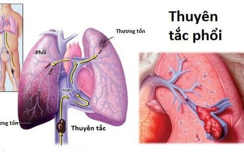
A pulmonary embolism occurs when a mass of material, usually a blood clot, enters an artery in a person's lungs. These blood clots commonly occur in deep vein thrombosis (DVT) in the deep veins of the patient's legs.In many cases, not one, but many blood clots can lead to a pulmonary embolism. If any part of the lung is blocked by an artery supplying blood, this is called a pulmonary infarction. This makes it harder for the lungs to supply enough oxygen to the rest of the body.
Sometimes, a blockage in a blood vessel is caused by substances other than a blood clot, such as:
Fat from broken bone marrow Part of the tumor Air bubbles
3. Diagnosis of acute pulmonary embolism
Pulmonary embolism can be difficult to diagnose, especially in people with underlying heart or lung disease. For this reason, the doctor will ask about the patient's medical history, do a physical exam, and perform one or more of the following diagnostic tests/imaging techniques:3.1 Blood tests Your doctor may order a D-dimer test to look for signs of increased clotting, although many other factors can also cause D-dimer levels to be high.

Xét nghiệm để tìm các nguyên nhân có thể làm tăng khả năng đông máu
3.3 Ultrasound (Ultrasound) Ultrasound uses sound waves to scan the veins in a person's thighs, knees, and calves and sometimes in the arm to check for blood clots in the deep veins.
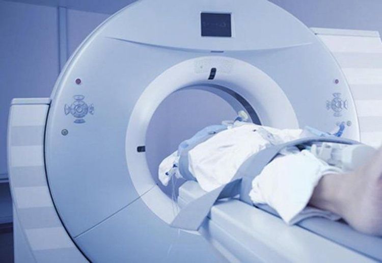
3.4 Computed tomography pulmonary angiography (CT pulmonary angiography) CT scans emit X-rays to create cross-sectional images of the patient's body. CT scans create 3D images to detect abnormalities such as an embolism in a pulmonary artery. In some cases, the patient may need to inject contrast material during the CT scan to make the pulmonary arteries clearer and easier to detect.

Chụp CT để chẩn đoán bệnh tắc mạch phổi
3.6 Pulmonary angiogram This technique gives a clear picture of the blood flow in the pulmonary arteries of the patient so it is the most accurate way to diagnose a pulmonary embolism. However, this technique requires a highly skilled technician and carries some risks, so it is usually performed only when other diagnostic techniques do not provide sufficient information. to accurately diagnose pulmonary embolism or not.
During a pulmonary angiogram, a flexible tube is inserted into a large vein - usually in your groin - and threaded through your heart into your pulmonary artery. A special dye is then injected into the catheter and X-rays are taken as the dye travels along the arteries in your lungs.
In some people, this procedure may cause a temporary change in heart rate. In addition, the dye may increase the risk of kidney damage in people with impaired kidney function.
3.7 Magnetic Resonance Imaging (MRI) This is an imaging technique that uses a computer-generated magnetic field and radio waves to create detailed images of a patient's organs and tissues. Magnetic resonance imaging is usually reserved for pregnant women to avoid radiation to the fetus and people with kidney disease to avoid the risk of further kidney damage due to the dyes used in these diagnostic techniques. above.
Currently, at Vinmec Times City International Hospital, fibrinolysis is being applied for emergency treatment of patients with pulmonary embolism. With modern imaging equipment, CT perfusion results are available within 7 minutes and performed routinely. The technique is performed by a team of qualified, experienced doctors and modern equipment and professional services.
Please dial HOTLINE for more information or register for an appointment HERE. Download MyVinmec app to make appointments faster and to manage your bookings easily.


