This is an automatically translated article.
The article was professionally consulted by Specialist Doctor II Mai Anh Kha - Orthopedic surgeon - Department of General Surgery - Vinmec Danang International General Hospital.A series of occupational accidents, daily-life accidents, sports accidents ... are increasing rapidly and accompanied by a sharp increase in the rate of maxillary fractures (in the group of maxillofacial injuries) in the country. ta. The diagnosis and treatment of maxillary fractures depends on the extent and nature of the injury.
1. Learn about the anatomy of the maxilla
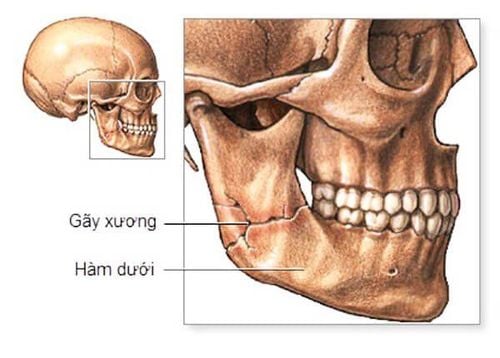
The maxillary bone is composed of two bones that are symmetrical through a central longitudinal plane, creating the midfacial mass. Therefore, when trauma occurs in the maxillary bone, it will often be accompanied by other injuries in the middle facial bones such as the main bone of the nose, cheekbones, lacrimal bone, cane bone, lower curly bone...Jawbone The upper jaw is also closely related to the eye socket, nasal cavity, skull base and maxillary sinus, so trauma to the maxillary bone also has serious effects on the sensory organs and the skull. The maxillary region is a fixed bone area, the upper part is covered by the base of the skull and the main bone of the nose, the two sides are the cheekbones, the temporal bone, and the lower part is the alveolar bone, the lower jaw bone. Therefore, fracture of the maxilla only occurs when strong and direct forces are applied.
At the same time, the maxillary bone is also a spongy bone, which is nourished by many blood vessels. Therefore, when a fracture occurs, the patient will bleed a lot, requiring emergency care.

2. How are maxillary fractures classified?
Fractures of the maxilla are divided into 2 main types.Partial fracture: Including cases such as fracture of the ascending process, fracture of the alveolar bone, fracture of the lower orbital bone, subsidence of the canine fossa, fracture of the apex,...
Total fracture
Severe case of jaw fracture above, including:
Longitudinal fracture. Horizontal fracture.
3. How to diagnose maxillary fracture?
3.1 Partial fracture of maxilla
When the patient has a partial maxillofacial trauma, the diagnosis will be based on:For maxillary fractures of the ascending branch:
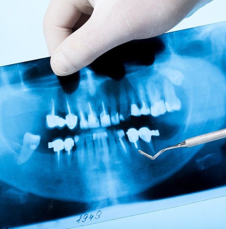
Bruising in the inner corner of the eye, sharp pain or slightly concave to the injury site. Nosebleed . Excessive tearing may occur because the patient has a blocked tear duct. Through X-ray or CT Scanner, the profile will show the image of the fracture line in the upper jaw area. For fracture of the anterior maxillary sinus:
Nosebleed. Bruising under the eye socket and possibly numbness of the affected cheek. Through inspection images, there is damage to the anterior wall of the sinus. For suborbital and orbital floor fractures:
Nose bleed. Concave eyes, signs of diplopia. Numbness in the cheeks, there is a sharp pain below the eye socket. X-ray or CT Scanner shows lesions below the orbit.
3.2 Total jaw fracture
Patients with total maxillofacial trauma will present with shock and accompanied by traumatic brain injury. Depending on the type of jaw fracture is vertical or horizontal, the patient will have different symptoms.Longitudinal maxillary fracture:
Clinically: the patient has nose and mouth bleeding, malocclusion, when examining the bones, the upper jaw is mobile. Image: using X-ray technique or CT Scanner, Belot maxillary, noticed the image of lesions along the middle or along the maxillary bone. Horizontal maxillary fracture:
Clinically: Lefort I fracture leads to upper lip bruising, malocclusion; Lefort II fracture leads to facial swelling, bilateral hematoma, fresh nose bleeding, malocclusion; Lefort III fracture leads to facial edema, bilateral orbital bruising, conjunctival hematoma, diplopia, palpable displaced bone ends. Image: X-ray or CT-Scanner shows transverse fracture.
4. How to treat upper jaw fracture?
Jaw fractures can come from many causes, which are mainly physical such as traffic accidents, accidents in daily life or work accidents... Therefore, it can lead to many different types of fractures. . Depending on the type of jaw fracture, the treatment will be indicated specifically.Principles of treatment
Doctors need to adhere to the principles of treatment of maxillary fractures including:
Correction of broken bones. Fix broken bones. Optimal prevention of complications from occurring. Need rehabilitation and aesthetic treatment for the patient. Some specific treatment of maxillary fracture
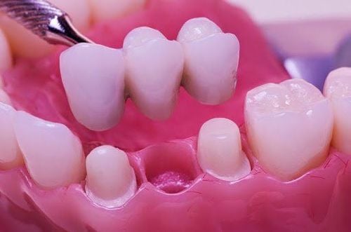
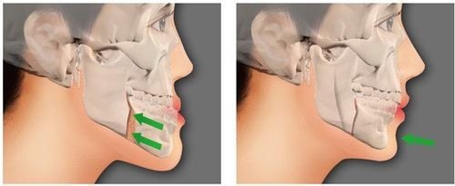
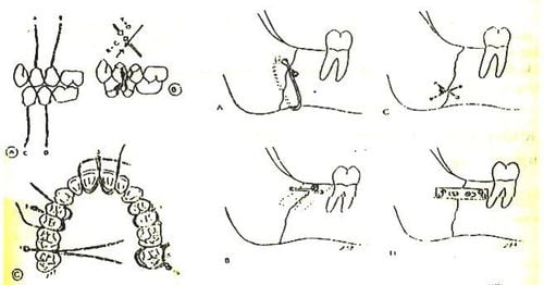
Maxillary suspension surgery. Surgery to combine jawbone with screw brace.
5. Prognosis and complications of maxillary fractures
Prognosis: Jaw fractures, especially maxillary trauma, can have serious consequences for patients' health and quality of life. In which:When being treated as soon as possible and with the right treatment principles, the patient will have a high chance of recovery. Late treatment or wrong treatment principles can cause serious complications, not only affecting aesthetics but also causing problems with living functions in the body. Possible risks of performing maxillary fractures: Infection. Misaligned bite. Mouth-opening activity is limited. A fracture of the upper jaw is a serious accident that needs to be treated as soon as possible. In addition, it is also necessary to take preventive measures such as preventing traffic accidents, providing good protection in working and living activities.
Currently, at Vinmec International General Hospital, the surgical method of jaw bone correction has been applied with the advantage of correcting malocclusion due to bone structure, returning the function of chewing, biting, and breathing well. cosmetic facial harmony and radical change in beauty.
Patients need to be examined soon after trauma for explanation and surgical options. Vinmec International General Hospital is the leading prestigious address with:
Good MRI machines Experienced specialists, sterile operating room, strict surgical procedures. Any questions that need to be answered by a specialist doctor as well as customers wishing to be examined and treated at Vinmec International General Hospital, please book an appointment on the website to be served.
Please dial HOTLINE for more information or register for an appointment HERE. Download MyVinmec app to make appointments faster and to manage your bookings easily.


