This is an automatically translated article.
The article is professionally consulted by Master, Doctor Pham Van Hung - Department of Medical Examination & Internal Medicine, Vinmec Danang International General Hospital.Atrioventricular block (AV block) is a disorder of conduction from the atria to the ventricles diagnosed by electrocardiograms, causing arrhythmias that can cause syncope or sudden death. Depending on the degree of block there are different treatment methods
1. What is atrioventricular block?
Atrioventricular block is a partial or complete obstruction of the conduction of impulses from the atria to the ventricles manifested by prolongation of the PR interval on the electrocardiogram. The most common cause is fibrosis or necrosis of the conduction system. The atrioventricular node conducts and controls electrical impulses from the atria to the ventricles. The sinus node generates conduction to the atria causing the atria to contract, as shown by the P wave on the electrocardiogram. It is then transmitted to the atrioventricular node, bundle of His, to the Purkinje network, causing the ventricles to contract, showing the QRS complex on the electrocardiogram. When the impulses from the atria to the ventricles are obstructed to varying degrees at the atrioventricular node or bundle of His, it is called atrioventricular block.Depending on the degree of obstruction, the conduction of impulses from the atria to the ventricles, atrioventricular block is divided into 3 degrees: atrioventricular block I, II, III.
2. Causes of atrioventricular block
There are many causes of atrioventricular block including:Exercise and sports Myocardial infarction Mitral valve replacement surgery, mitral valve repair Myocarditis Idiopathic fibrosis of the conduction system in Lenegre disease or Lev disease autoimmune ( lupus erythematosus , systemic sclerosis ). Hyperkalemia. Due to drugs such as beta-blockers, calcium channel blockers, digoxin, amiodarone.
3. Diagnosis of atrioventricular block
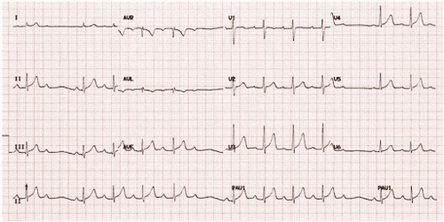
Diagnosis of complete atrioventricular block is based on electrocardiogram test3.1 For first-degree atrioventricular block Diagnosed when the ECG has prolonged PR interval > 200ms equivalent to 1 large cell (Figure 1), the diagnosis is clear. when PR interval >300ms and/or P wave is hidden in previous T waves (Figure 1, Figure 2)


For second-degree atrioventricular block type 1: PR interval gradually lengthens, eventually a P wave without QRS. Longest PR interval just before skipping beat. The PR interval is the shortest immediately after skipping (Figure 3).


4. Treatment of atrioventricular block
There is no specific treatment for patients with atrioventricular block. Patients need to be hospitalized for treatment when there are symptoms of shortness of breath, syncope, chest pain... In addition, the patient should be controlled for pulse and blood pressure with vasopressors or blood pressure control drugs. Specifically as follows:For first-degree atrioventricular block:
Characteristics that do not cause hemodynamic disturbances. No specific treatment is needed. Some cases of first-degree atrioventricular block with right bundle branch block may increase the risk of progression to third-degree atrioventricular block, so it is necessary to closely monitor the pacemaker for second-degree atrioventricular block Mobitz I
Especially The score is usually a benign arrhythmia, causing minimal hemodynamic instability and low risk of progression to third-degree atrioventricular (AV) block. Asymptomatic patients do not require treatment. Permanent pacing is rarely needed. For second-degree atrioventricular block Mobitz II
Features more hemodynamic instability than Mobitz I, severe bradycardia and progression to third-degree atrioventricular (AV) block, which can cause syncope or sudden death. Mobitz II heart failure requires immediate hospitalization for cardiac monitoring, temporary pacing, and ultimately, a permanent pacemaker. For third-degree atrioventricular block
Characteristics: Patients with third-degree atrioventricular (AV) block are at high risk of ventricular arrest and sudden cardiac death. Urgent hospitalization is required for cardiac monitoring, temporary pacing, and often permanent pacemaker placement. To protect cardiovascular health in general and detect early signs of cardiovascular disease, customers can sign up for Cardiovascular Screening Package - Basic Cardiovascular Examination of Vinmec International General Hospital. The examination package helps to detect cardiovascular problems at the earliest through tests and modern imaging methods. The package is for all ages, genders and is especially essential for people with risk factors for cardiovascular disease.
To receive examination and treatment with leading Vinmec cardiologists, please book an appointment online at the website or contact Vinmec Health System nationwide for service.
Please dial HOTLINE for more information or register for an appointment HERE. Download MyVinmec app to make appointments faster and to manage your bookings easily.


