This is an automatically translated article.
Ankylosing spondylitis is a type of birth defect associated with the skull of an infant being deformed, thereby causing abnormalities of the brain. The method to improve the condition of the skull stenosis in the newborn is being researched and applied more and more widely nowadays, is cranial plastic surgery, which helps the child's brain to develop and reverse the consequences. serious consequences of this disease.
1. Ankylosing spondylitis
The skull is an organ in the human body that protects the brain, including bones such as the ethmoid, frontal, occipital, parietal, sphenoid, and temporal bones. In an individual, when developing normally, these skull bones will tend to separate when the child reaches 2 years of age, interlock when the child is 2 to 4 years old, and fuse after the age of 20. Craniosynostosis or craniosynostosis, scientifically known as Craniosynostosis, is a type of birth defect in children caused by the fusion of the skull joints right from birth.Ankylosing spondylitis causes the skulls of children to become deformed, narrower than those of normal-born children. Usually craniocervical stenosis will appear during the fetal period in the womb, the manifestation of the disease if it occurs during this period is that the baby will be born with an abnormality in the shape of the head and face. In addition, craniosynostosis also inhibits the growth of the skull, leading to increased intracranial pressure in children, causing symptoms such as headaches, visual disturbances and growth retardation.
The disease has many different degrees, each disease form will have some different pathological features and depend on the type of joint involved as well as the time when the child has ankylosing spondylitis. However, for a baby born with a deformed head, it is not possible to confirm with certainty that the cause is ankylosing spondylitis because it can be the effect of some problems such as the fetal head position during pregnancy. in the womb or suffered trauma during childbirth. After a while, skull abnormalities due to the above causes can be completely treated, but if the cause is an ankylosing spondylitis, the cure rate will be very low if not treated promptly.

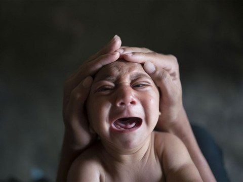
Dị tật dính khớp sọ thường xảy ra khi trẻ còn là bào thai
Clinical forms of craniocervical malformations include:
One-sided coronal fusion: From the ear to the longitudinal joint, the clinical manifestation is anterior distortion of the head, flattened forehead on one side, and an anterior orbit of the eye socket upward. , skull and nose deviated to one side, vision loss, blindness... Lamda fusion: Very rare joint in craniosynostosis cases, manifests as posterior deformity, protrusion of mastoid and posterior deviation of ears. , the skull is distorted to one side... Adhesion of the coronal joint line on both sides: The coronary joints are fused on both sides, leading to some manifestations such as short, wide head, flat forehead and eyebrows, raised and sunken. Adhesion of the frontal joint line: From the nose to the longitudinal joint, causing triangular head, pointed forehead, high ridge of bone in the middle of the forehead, the eyes are very close... Adhesion of the longitudinal joint line: Is a form of sticky malformation Cranial joints are most common, causing a boat-shaped head, protruding forehead, and head that is elongated anteriorly but narrowly transversely.
2. Cranioplasty in patients with craniocervical stenosis
Narrow skull defects can lead to increased intracranial pressure in children, causing brain damage. Therefore, when children are found to have cranial ankylosing spondylitis, they need cranioplasty as soon as possible so that their brains can develop normally, prevent intellectual disability or a number of diseases related to the brain. The best recommended time to carry out cranial shaping for children is under 12 months of age, more specifically, between 3 and 8 months of age, the most optimal treatment effect can be achieved.
To be able to shape the skull, it is necessary to prepare a number of problems such as:
Diagnosis of the disease by examination, X-ray, computed tomography... to detect stenosis of the skull as well as some diseases other reasons attached. Clean the head, shave the hair before surgery. Prepare the required amount of blood before surgery. Fully equipped with necessary tools including pediatric craniofacial instruments, cranial microsurgery instruments, microsurgery glasses, craniotomy drill, skull pins, specialized pediatric screws with the ability to dissolve.. .

Chẩn đoán bằng hình ảnh trước khi phẫu thuật (hình ảnh minh họa)
Steps to perform skull shaping include:
Intubation anesthesia for children. Make two incisions from the baby's temple, side to side of the temple to peel off the scalp. Exposing the fused joint on the skull. Use drill to drill bone and cut adhesive joints as well as damaged bones if any. Shaping the skull, which can be part or all of the skull. Use self-dissolving screws and self-dissolving splints to fix the skull bones. Hemostasis, drainage under the scalp. Suture the scalp and compression bandages as well as wear a helmet after surgery. After surgery, the patient needs to be monitored for a number of important issues such as:
Vital signs include pulse, breathing rate, temperature and blood pressure. Take prophylactic antibiotics and pain relievers. Note the focal neurological signs to timely detect neurological complications. If there is bleeding after surgery, it is necessary to monitor and stop bleeding according to the degree of bleeding. When the patient has an infection or meningitis, it is necessary to culture blood, culture cerebrospinal fluid to make an antibiotic map for effective antibiotic treatment. Some children after cranioplasty may experience hydrocephalus, which can be treated initially by draining the ventricles, then monitored and treated. Ankylosing spondylitis is a congenital malformation in infants at birth, caused by early fusion of some skull joints leading to a condition where the brain cannot develop normally. The disease affects children's intellectual and neurological development, making it difficult for children's health, growth and development as well as daily activities. However, if the condition of craniocervical stenosis is detected in time and treated with cranioplasty in an early time, the child is completely capable of normal brain development, learning, functioning and development. good after successful treatment.
Vinmec International General Hospital with a system of modern facilities, medical equipment and a team of experts and doctors with many years of experience in neurological examination and treatment, patients can completely peace of mind for examination and treatment at the Hospital.
To register for examination and treatment at Vinmec International General Hospital, you can contact Vinmec Health System nationwide, or register online HERE.
LEARN MORE
Causes of neonatal cranial ankylosing spondylitis in children Common types of craniosynostosis in children Diagnosis and treatment of early craniosynostosis in children