This is an automatically translated article.
The article is professionally consulted by Master, Doctor Vu Huy Hoang - Radiologist - Department of Diagnostic Imaging and Nuclear Medicine - Vinmec Times City International General Hospital.Sinusitis is a common disease in Vietnam due to many causes, the most common being environmental pollution. The goal of computed tomography scanning of the sinuses is to determine whether facial pain is caused by sinus disease or other causes, to assess the status and extent of the disease, and to support treatment.
1. Anatomy of sinusitis
Modern medicine believes that inflammation is a biological process including many reactions of the body, local and systemic, to protect the body against pathogens, repair damaged areas to re-establish. homeostasis. The inflammatory process consists of many different stages and phenomena. Inflammatory factors can be from the external environment or form within the body.Phenomena through inflammatory stages:
Initiation (biochemical phase): Occurs early in the first few days, difficult to recognize clinically, includes many chemical and biological reactions. The vascular - blood reaction includes two periods: the early period (I), which is a few minutes long, due to the action of histamine; The late period (II), due to the action of kinins, reaches a maximum within 3 - 4 hours. During this phase, transient vasoconstriction occurs first, followed by hemodynamically congestive vasodilatation. Based on clinical progression, there are 3 classifications of inflammation:
Acute inflammation: Lasts a few days to a few weeks, including many vascular - blood reactions with infiltration of many nuclei. Subacute inflammation: Lasts several weeks to several months, consisting of tissue reaction with inflammatory granulomatous tissue present. Chronic inflammation: Lasts many months, many years, including healing and repair reactions, accompanied by fibrous hyperplasia and fibrosis. Otolaryngology in general and sinusitis in particular are often divided into two types, acute inflammation and chronic inflammation, from which to decide on medical treatment or surgery. There are also polypoid lesions, which can be isolated or associated with chronic inflammation.
2. Chronic sinusitis
Chronic rhinosinusitis is a condition in which clinical symptoms persist for more than 12 weeks. This is one of the most common chronic diseases, accounting for a high proportion of ear - nose - throat problems. According to statistics, the rate of chronic rhinosinusitis in the community accounts for 14% of the population.Previously, the diagnosis of rhinosinusitis was difficult and inconsistent. But with the development of science and technology, especially CT scanning of the sinuses, the diagnosis is not only easier, but also effectively supports the endoscopic sinus surgery method. At present, it is possible to identify the lesion, evaluate the degree of ventilation in the complex of the nook and cranny, and other abnormalities, etc., thereby preventing and limiting surgical complications.
The cause of sinusitis originating from the nose and often relapsing is due to changes in the niche complex. The fissure complex is located in the middle nasal cavity, changes here are factors that cause acute and chronic sinusitis. Computed tomography scan of the sinuses with two positions Axial and Coronal helps to assess the thickness, opacity of the sinuses, complex of holes... as well as the degree of pathology according to the Lund - Mackay scale.
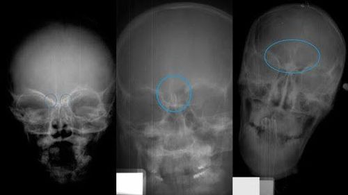
Chụp CT khảo sát xoang giúp việc chẩn đoán bệnh dễ dàng hơn
3. Computed tomography scan of the sinuses
In 1972, a British engineer and American doctor built a complete CT scanner. By 1995, many radiologists had recognized the role and value of CT scan in the diagnosis of sinusitis. Specifically, it helps to provide otolaryngologists with accurate information on:Anatomy of complex slit openings Signs of sinus disease: Nasal polyps, crooked nasal septum, thickening of mucous membranes in the sinuses ... Abnormal changes in the structure of the nasopharynx, in the ethmoid and sphenoidal sinuses. Size of Agger nasi cells, frontal recess, orbit, ethmoid ball, ethmoid ceiling, posterior ethmoid cells, ethmoid funnel ... Abnormal septum septum in sphenoid sinus Congenital lack of bone wall causes carotid artery to be exposed in sphenoid sinus CT scan is indicated when:
Sinus disease has been treated with maximum medical treatment but still fails Failure Preoperative evaluation of endoscopic sinusitis Complicated sinusitis Suspected malignancy, naso-sinus tumor. Computed tomography of the sinuses is usually done through 2 planes:
Frontal (Coronal) plane: The patient lies supine or prone, the neck is maximally supine. The cross-section is perpendicular to the hard palate. Horizontal plane (Axial): The patient lies on his back, head straight, eyes looking up at the ceiling. The cross-section is parallel to the palate. Today, many medical professionals recommend CT scans in the coronal position to examine the sinuses more clearly than in the horizontal plane. The frontal plane CT scan shows:
Sequential anatomical structures from front to back as when performing surgery Relation of the sinuses to important adjacent anatomical structures, such as the ceiling of the ethmoid sinus, second bone Image is more than 100 times clearer than conventional X-rays, clearly showing the anatomical structure of the nasal cavity, the sinus ostomy complex, the sinuses and related structures Special pathological images of the sinuses such as: maxillary sinus insufficiency, concha bullosa, cystic fibrosis, Aspergillus cysts in the sinuses. Therefore, CT scan coronal position is the most suitable to approach the nasopharyngeal lesions, considered the gold standard to evaluate the status of sinusitis. and structural abnormalities of the sinuses.
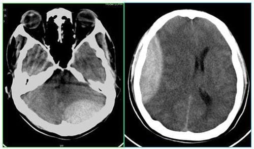
CT scan tư thế coronal được xem là chuẩn vàng để đánh giá tình trạng viêm xoang
4. A CT scan of the sinuses to assess the extent
To evaluate severe or mild chronic sinusitis on CT scan, many doctors use a scale based on the opaque images of the sinuses and the cleft palate complex to score, specifically as follows:| Hình ảnh mờ đục | Bên phải | Bên trái |
|---|---|---|
| Xoang hàm | 0 - 2 | 0 - 2 |
| Xoang sàng trước | 0 - 2 | 0 - 2 |
| Xoang sàng sau | 0 - 2 | 0 - 2 |
| Xoang bướm | 0 - 2 | 0 - 2 |
| Xoang trán | 0 - 2 | 0 - 2 |
| Phức hợp lỗ thông khe | 0 - 2 | 0 - 2 |
| Tổng cộng | 12 | 12 |
0: no abnormalities 1: partial opacity 2: complete opacity. For slit opening:
0: no obstruction 1: partial obstruction 2: complete obstruction In summary, patients with sinusitis should be assigned a CT scan of the nasal sinuses. In addition to determining the correct sinusitis and extent, CT scan images also help evaluate the complex malformation of the foramen, septum deformity, and mid-slit malformation. It is necessary to combine clinical with endoscopy and computed tomography to investigate the sinuses to improve efficiency in the diagnosis and treatment of sinus disease.
Vinmec International General Hospital is one of the hospitals that not only ensures professional quality with a team of leading medical doctors, a system of modern equipment and technology. The hospital provides comprehensive and professional medical examination, consultation and treatment services, with a civilized, polite, safe and sterile medical examination and treatment space. Customers when choosing to perform diagnostic imaging here can be completely assured of the accuracy of the results.
Please dial HOTLINE for more information or register for an appointment HERE. Download MyVinmec app to make appointments faster and to manage your bookings easily.
Reference sources: benhviemxoang.vn, soyte.hatinh.gov.vn