This is an automatically translated article.
The article is professionally consulted by Master, Doctor Nguyen Le Thao Tram - Radiologist - Radiology Department - Vinmec Nha Trang International General Hospital.Computed tomography brain angiography is one of the optimal methods for timely detection of cerebral infarction and cerebral hemorrhage. It is also an effective support tool to help doctors quantify cerebral vascular occlusion by region. In addition, CT angiography of the brain will also evaluate lesions such as cerebral aneurysms, cerebral artery malformations... early diagnosis of cerebral stroke.
1. What is a CT angiogram of the brain?
Brain stroke, also known as cerebrovascular accident, is a very common disease in the emergency room. It is divided into two types namely cerebral infarction (cerebral blood vessel occlusion) and cerebral hemorrhage (cerebral bleeding). The disease often occurs in the elderly with other diseases such as diabetes, hypertension, dyslipidemia, cerebral aneurysms... Therefore, detecting signs for diagnosis and treatment regimens Timely treatment of these patients will save their lives and reduce the rate of disability or sequelae left by the disease.
To diagnose the disease accurately and early, the method of cerebral vascular computed tomography will be one of the preferred choices of doctors. This is one of the methods using advanced imaging techniques along with X-rays to diagnose neurovascular diseases.
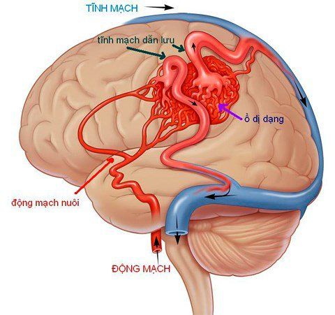
CT cerebral angiography is capable of diagnosing cerebrovascular malformations, cerebral aneurysms, cavernous carotid artery fistulas or cases requiring evaluation for stenosis, cerebral artery thrombosis, and venous sinuses. circuit.
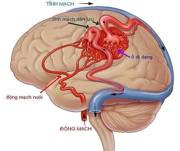
Chụp cắt lớp vi tính giúp phát hiện những dị dạng mạch máu não
2. When is it indicated and contraindicated in what cases?
2.1. Indication for CT angiography of the brain
The doctor will order a CT angiogram of the brain in the following cases:
● People who suspect brain vessel abnormalities such as subarachnoid hemorrhage, bleeding brain parenchymal bleeding, intraventricular bleeding...
● People with signs of cerebrovascular malformation, or epilepsy due to cerebral vascular malformation
● People with stroke due to cerebral infarction, arterial infarction , venous infarction
● People with arteriovenous fistula, cavernous sinus fistula
People with venous thromboembolism, cerebral venous sinus
● People with skin vascular malformations head
● People with meningiomas and need to evaluate the tumor's vascular source...
In addition, this method is also used for monitoring after treatment of cerebrovascular disease. For cases requiring surgical intervention or embolization, it is necessary to require tomography with 64 sequences or more to evaluate the entire area of metal interference.
2.2. Contraindications to CT angiography
Cases in which the examination area (cranial) contains many metals that interfere with the test results
Pregnant women
Reactions allergic to iodinated contrast agents.
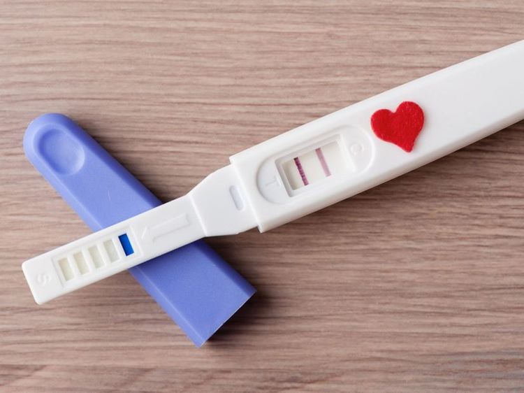
Phụ nữ có thai không được chụp CT mạch máu não
3. What does the patient need to prepare before the scan?
Before the CT scan, the patient will be asked to remove all metal objects such as jewelry, dentures, hairpins... Because they may affect the test results.
The patient will be asked not to eat or drink anything for several hours (about 4 to 6 hours) before the test.
The patient will be explained in detail by the specialist and the technician to help the patient cooperate more easily.
If the patient cannot lie still, the doctor may ask the patient to use sedation.
4. Steps to perform cerebral vascular computed tomography
The patient is instructed to lie on the table of the machine, in a relaxed position. At the same time, the patient will be secured with a belt so that the correct position can be maintained during the examination. If the patient has small movements, it will make the image blurry, reducing the quality of the image as well as the test results.
The technician starts positioning and sets the brain scan field. The next step is to emit X-rays and process images to evaluate brain parenchyma, and at the same time select the necessary images that can observe the pathology most clearly.
Carry out intravenous placement with an 18G needle, connect a double-bore electric injection pump (in which one is drug and one is physiological saline) with iodinated contrast. First, a non-contrast scan will be taken. The results were obtained to perform a bolus test of the common carotid artery at the level of the C4 cervical vertebra. Then, inject contrast and continue to use X-rays to take pictures. The acquired images will be systematically visualized of the cerebral arteries to highlight the pathology related to the cerebral blood vessels.
CT cerebral angiography is capable of diagnosing cerebrovascular malformations, cerebral aneurysms, cavernous carotid artery fistulas or cases requiring evaluation for stenosis, cerebral artery thrombosis, and venous sinuses. heart disease and many other diseases. In fact, brain diseases, if not examined and treated early, can leave dangerous complications, even lifelong sequelae.
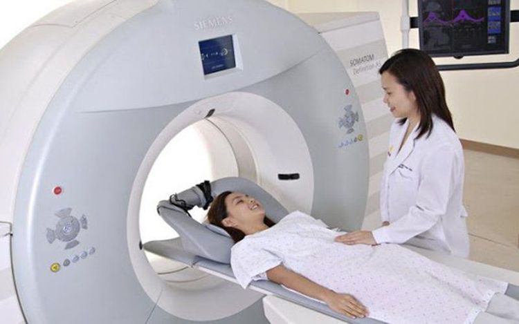
Bệnh nhân thực hiện chụp cắt lớp theo hướng dẫn của nhân viên y tế
Computed tomography (CT) of cerebral blood vessels is one of the imaging techniques applied by Vinmec International General Hospital, from which to detect brain diseases, cerebrovascular malformations,... early diagnosis of cerebral stroke at an early stage,... Especially to improve diagnostic results, Vinmec has been and continues to invest in modern CT, X-ray, and MRI machines. .. combined with medical services of examination, treatment and early screening of cancers.
Especially now, Magnetic Resonance Imaging - MRI/MRA is considered a "golden" tool for brain stroke screening. MRI is used to check the condition of most organs in the body, especially valuable in detailed imaging of the brain or spinal nerves. Due to the good contrast and resolution, MRI images allow to detect abnormalities hidden behind bone layers that are difficult to recognize with other imaging methods. MRI can give more accurate results than X-ray techniques (except for DSA angiography) in diagnosing brain diseases, cardiovascular diseases, strokes,... Moreover, the process MRI scans do not cause side effects like X-rays or computed tomography (CT) scans.\
Vinmec International General Hospital currently owns a 3.0 Tesla MRI system equipped with state-of-the-art equipment by GE. Healthcare (USA) with high image quality, allows comprehensive assessment, does not miss the injury but reduces the time taken to take pictures. Silent technology helps to reduce noise, create comfort and reduce stress for the client during the shooting process, resulting in better image quality and shorter imaging time. With the state-of-the-art MRI system With the application of modern methods of cerebral vascular intervention, a team of experienced and well-trained neurologists and radiologists, Vinmec is a prestigious address for stroke risk screening and screening. reliable goods.
In the past time; Vinmec has successfully treated many cases of stroke in a timely manner, leaving no sequelae: saving the life of a patient suffering from 2 consecutive strokes; Responding to foreign female tourists to escape the "death door" of a stroke;...


