
This is an automatically translated article.
The article is professionally consulted by MSc, BS. Dang Manh Cuong - Doctor of Radiology - Department of Diagnostic Imaging - Vinmec Central Park International General Hospital. The doctor has over 18 years of experience in the field of ultrasound - diagnostic imaging.Pancreatic trauma can result from penetrating trauma or perforation of the abdomen with a very rare but high mortality rate. In this context, computed tomography of pancreatic trauma is the preferred imaging modality, thereby directing timely intervention for patients.
1. What is pancreatic injury?
Due to the deep anatomical location in the abdomen, anterior to the spine, pancreatic trauma is rare and relatively difficult to diagnose, so the diagnosis can often be missed. In contrast, injuries to the liver, spleen, and kidney are common and often easily identified by conventional imaging modalities.The first step to avoid missing pancreatic injury is to consider the mechanism of injury and then evaluate the anatomical structures involved. Severe anterior-posterior abdominal trauma, which compresses the pancreas into the spine, such as seat belt injuries, deceleration injuries, and handlebar compression injuries are the most common mechanisms of injury. . Now, in suspected cases, pancreatic injury is often identified by various imaging modalities, notably by pancreatic tomography, and also to investigate associated multi-organ injuries.
The most obvious signs of pancreatic injury due to the mechanism of pancreatic injury are pancreatitis with blood spread around the pancreatic capsule, edema, and soft tissue infiltration extending to the pre-adrenal region. Changes in post-traumatic pancreatitis may not be detected for several hours after injury because they are time-dependent. Therefore, multiple CT scans of repeat pancreatic injury are needed, because delayed diagnoses of pancreatic injury are associated with high later mortality.
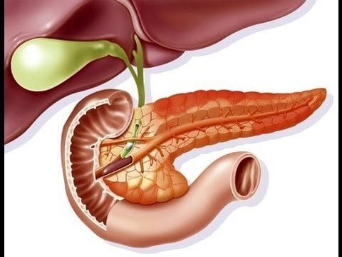
Chấn thương tụy tạng là hiếm gặp và tương đối khó chẩn đoán nên thường bị bỏ sót
2. Pancreatic injury image on pancreatic tomography
Multi-slice computed tomography is a useful imaging modality in patients with severe trauma and, in particular, it is the preferred imaging modality of choice for detecting acute pancreatic injury with high sensitivity. However, the sensitivity of pancreatography can still be variable because the ability to image discreet pancreatic lesions within the first 12 hours after injury is sometimes difficult. This is a consequence of the small amount of peripancreatic adipose tissue that limits the detection of pancreatic injury or the early admission of patients to the hospital, the lack of time required for the development of imaging manifestations of inflammatory phenomena following pancreatic trauma. .Because the presence of pancreatic lesions affects treatment, the disease status needs to be accurately determined. At this time, besides computed tomography of pancreatic trauma, the doctor can combine magnetic resonance imaging or endoscopic retrograde cholangiopancreatography.
Signs of pancreatic injury that may be suspected indirectly or directly described by computed tomography of pancreatic injury are as follows:
Indirect signs are simulations of the direction of force in the lesion. injury to the pancreatic region and the presence of peripancreatic fat fibers. Direct findings on CT scans of the pancreas can range from shrinkage of pancreatic tissue, edema or hematoma in the parenchyma, to a tear or rupture of the pancreas: Pancreatic edema is an area of low density on film, to showed edematous infiltration of the pancreatic parenchyma due to post-traumatic inflammation. Hematomas are also hypoattenuated areas, representing any amount of blood that may be confined to the parenchyma or may be extensive in the peripancreatic tissue. Furthermore, in severe cases, it may be associated with massive bleeding from the pancreas into the abdomen due to damage to the blood vessels in the pancreas. If the hematoma is large, it can also cause a mass effect to block the lumen of the duodenum adjacent to the pancreas or cause compression of the pancreatic duct.

Chụp cắt lớp được ưu tiên lựa chọn phát hiện tổn thương tụy cấp với độ nhạy cao
In addition, when the pancreas is involved in abdominal trauma, it is especially important to carefully evaluate the duodenum on CT because this organ can also be injured along with the pancreas. Imaging findings of duodenal involvement may not be obvious, but thickening of the duodenal wall or the presence of small air bubbles in the extragastric space into the vicinity may be indicative of injury. accompanying duodenal injury.
3. Complications of pancreatic injury on pancreatography
Complications following pancreatic trauma may occur in up to one-third of patients, including pancreatitis, pancreatic pseudocyst, fistula, intra-abdominal abscess, or pancreatic duct rupture. These complications can lead to a severe outcome of sepsis and/or multiple organ failure, and ultimately death.
Chấn thương tụy không được điều trị kịp thời dễ gây ra các biến chứng nặng nề
In summary, pancreatic injury resulting from blunt abdominal trauma or pancreatic injury is generally rare and difficult to diagnose. However, today's advanced imaging facilities, represented by computed tomography of the pancreas, allow for a complete and rapid assessment of the patient's condition. At the same time, doctors also need to understand the sensitivity of this tool to have a way to monitor patients with pancreatic injury appropriately, initiate early intervention and prevent consequences. However, to be able to accurately diagnose and treat effectively, patients need to perform computed tomography at a medical facility with a system of modern and prestigious equipment.
Currently, CT is an imaging method performed routinely at Vinmec International General Hospital with outstanding advantages such as: Equipped with many modern CT scanners such as 512 ranges, 640 ranges, State-of-the-art dual-energy CT, providing clear images of the body with high accuracy. They have the potential to help doctors eliminate the need for follow-up tests, while also helping patients shorten hospital stays. In addition, this technology also integrates a shield to regulate radiation exposure. At the same time, it also prevents unnecessary radiation doses before and after the procedure. With 640-sequence CT scanning technology, the time to record images of the coronary system is only in one beat, significantly reducing image noise caused by movement, so patients can save a lot of time for the examination process. mine. In particular, the computerized tomography procedure at Vinmec is carried out methodically under the guidance of the medical team of the Department of Diagnostic Imaging, which will help accurately diagnose the disease and the stage of the disease, thereby providing treatment. effective, creating a sense of safety for the patient.
Please dial HOTLINE for more information or register for an appointment HERE. Download MyVinmec app to make appointments faster and to manage your bookings easily.