This is an automatically translated article.
The article is expertly consulted by Master, Doctor Nguyen Hong Hai - Doctor of Radiology - Department of Diagnostic Imaging and Nuclear Medicine - Vinmec Times City International General Hospital.A colonoscopy is a technique that uses X-rays through a scanner to visualize the structure of the colon. Colonography scanning technique gives clear and detailed images. There is also the ability to visualize the colon from cross-sections, hence the name virtual colonoscopy.
1. Overview of CT Colonography
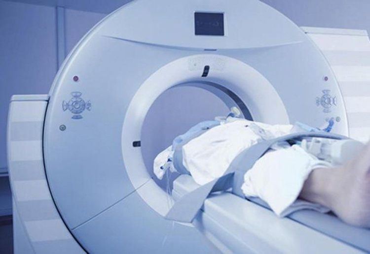
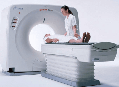
Computerized tomography (CT) is an imaging technique that uses high-intensity x-rays to scan areas of the body. A CT colonography or computed tomography colonoscopy is a multi-slice machine used to look at images of the rectum and colon or large intestine. Unlike colonoscopy, colonoscopy is a non-invasive method, but the images and information are similar. For this reason, a colonoscopy is also called a virtual colonoscopy.The multi-slice computed tomography machine consists of many parts, the most important of which is the X-ray chamber which is designed in the form of a giant round block. The patient lies on the x-ray table and is pushed into the imaging chamber with the part to be examined under the scanner emitting X-rays. The scanner inside the x-ray chamber will rotate continuously at high speed during the production process. X-ray beam. Different parts of the body with different densities corresponding to each type of tissue will have different density and reproduce images with different colors from white to gray to black.

Chụp cắt lớp đại tràng còn có tên gọi là nội soi đại tràng ảo
If you are over 50 years old, admitted to the hospital because of bloody stools, disordered bowel habits, weight loss, pale skin and mucous membranes, the doctor who suspects a colorectal malignancy may be diagnosed with colorectal cancer. to undergo a colonoscopy.
In addition, a prominent advantage of colonoscopy is that it is safe and minimally invasive, so colonoscopy can be indicated as an alternative to colonoscopy in patients with colonoscopy. elderly or frail patients with chronic medical conditions.
2. The procedure for performing a colon scan
Although CT colonography is quite safe and has few side effects, it needs to be performed correctly to provide good image quality as well as create comfort for the patient during the procedure. In general, each medical facility will have its own regulations on performing a colonoscopy, but still must follow the following main steps:Use muscle relaxants to reduce colon spasms to facilitate the get a good picture. Contrast-enhanced tomography may also be indicated. In this case, the patient will be prescribed oral contrast a few days before the procedure. Instruct the patient to lie on the table in a supine, supine, or prone position. Using straps to fix the knee and two legs to help the patient fix the posture during the shooting process, avoid image noise.

Quy trình thực hiện chụp cắt lớp đại tràng

Sau khi chụp CT đại tràng, người bệnh có thể xuất hiện cảm giác chướng bụng
3. What preparation is needed before a colonoscopy?
As a rule, patients will be guided and advised on what to do before performing a colonoscopy to ensure good image quality and safety of the technique. Some things to keep in mind include:Adhere to a special diet for a few days before the procedure Before a colonoscopy, enema or laxatives are needed to clear the bowel. Laxatives can make people tired in some cases as a side effect of diarrhea. With the indication for a colonoscopy with contrast, the patient will be instructed to take contrast two days before the scan. The main role of the contrast agent is to create a contrast that helps to create a more accurate image of the colon. In some cases, contrast media may be administered intravenously. At that time, the patient needs to be examined more closely to rule out contraindications and sometimes have to stop using some of the previous drugs.

Trước khi chụp cắt lớp đại tràng, người bệnh cần uống thuốc nhuận tràng để làm sạch lòng ruột
At Vinmec International General Hospital, the CT colonography procedure is performed by a team of highly qualified and experienced doctors in the field of diagnostic imaging, along with equipment, Modern machines for accurate and fast results.
Master, Doctor Nguyen Hong Hai graduated with a Master's degree in Diagnostic Imaging at Hanoi Medical University with strengths in diagnosing breast and thyroid cancer. Currently, Dr. Hai is working at the Department of Diagnostic Imaging, Vinmec Times City International Hospital.
To register for examination and treatment at Vinmec International General Hospital, you can contact the nationwide Vinmec Health System Hotline, or register online HERE


