This is an automatically translated article.
Chest trauma accounts for 20-25% of all trauma deaths. Chest trauma includes many different injuries such as contusion of the lung, pneumothorax, rib fracture, hemopericardium...
1. Chest trauma and causes
Chest trauma includes closed chest trauma and open chest trauma:
Closed chest trauma is trauma to the chest wall that is still closed, the pleural cavity is not open to outside air. Open chest trauma is an injury that causes a perforation of the thoracic cavity to open the pleural space to the outside air. The most common causes of thoracic injuries are traffic accidents, falls from above, accidents at work, injuries in sports practice, etc. Or a penetrating object such as: knives, scissors, bullets, iron bars...
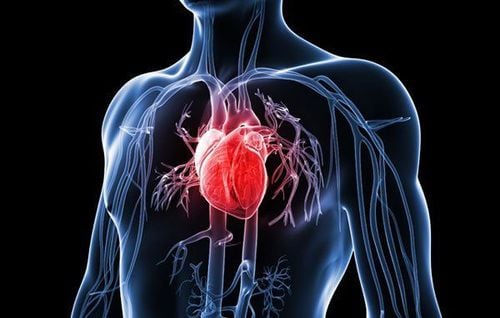
Thoracic trauma is extremely dangerous because it directly affects the body's circulatory system, so the mortality rate is high if not treated promptly.
2. Common injuries in chest trauma and treatment

Gãy xương sườn thường gặp trong chấn thương ngực
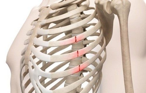
2.1 Rib Fracture Rib fracture is the most common injury in thoracic trauma, including two fracture mechanisms:
Direct fracture: That is, where the injury is broken there, direct rib fracture usually causes damage. lung injury. Indirect Fracture: When the ribcage is compressed, the ribs are easily broken from the inside out, causing damage to the heart and major blood vessels. Rib fractures can break one or more bones, causing the intercostal artery to rupture, causing heavy bleeding. In addition, rib fractures are sometimes associated with a sternum fracture.
Treatment of rib fractures by two methods:
Using large adhesive tape to fix the ribs, this is a simple and easy method to perform, but must be noted with elderly patients and patients with a history of injury lungs because this method will make the patient's breathing limited Intercostal nerve block: This method has many complications when injecting the intercostal artery, so it is no longer used In addition, the doctor will instruct the patient how to breathe so that the patient can inhale and exhale as much as possible, practice coughing and patting the patient to remove phlegm.
2.2 Mobile rib array This injury is common when the patient is directly traumatized, causing many respiratory and circulatory disorders.
Lateral costal plate is the most common site of this injury, it can be seen directly in clinical practice, in addition, it can also be seen in the sternum and posterior lateral ribs.
Treatment of movable rib array by external fixation of the rib plate with Judet brace and internal fixation with Rush nail through the fixed rib array above and below the one-bone fracture, mechanical ventilation intubation with muscle relaxation.
2.3 Lung contusion A lung contusion is a bruise of the lung that is the result of a blunt trauma to the chest area or caused by a high velocity fire. Pulmonary contusion causes hypoxia, which is aggravated if more lung tissue is congested.
Respiratory failure, hemoptysis are clinical signs of patients with pulmonary contusion.
Treatment of contusion is similar to that of mobile rib arrays, i.e. adequate analgesia, oxygenation and airway ventilation are the mainstays in the treatment of contusion. In addition, large volumes of fluid must be infused for cardiovascular resuscitation, but fluid overload should be avoided to avoid the development of ARDS syndrome.
2.4 Lung Tear Lung tear often causes large amount of blood loss because the blood flow through the lungs is very large and when the pulmonary blood vessels are damaged, it is difficult to stop bleeding. Patients with pulmonary lacerations require immediate surgery if the amount of blood through the drain exceeds 1500 ml after placement of the drain.
There are three methods to manage a lung tear: suturing for small or shallow lacerations, partial resection of the lung, and parenchymal resection to stop bleeding for deep lacerations.
2.5 Pneumothorax Pneumothorax in blunt chest trauma is caused by a broken rib impinging on the lung or from a tear in the lung. Note that tension pneumothorax will make the patient severely short of breath, requiring immediate first aid.
Pneumothorax is treated with continuous pleural drainage, for pneumothorax under pressure need needle aspiration of the pleural space to decompress before draining.

Tràn khí màng phổi
2.6 Hemothorax This is a very common complication in thoracic trauma. Blood can flow out from many different sources such as chest wall, lung, mediastinal blood vessels...
Depending on the amount of blood lost in the pleural space, the patient has symptoms such as chest pain, shortness of breath, pale skin, pale mucous membranes, rapid pulse and decreased blood pressure.
Treatment of hemothorax by aspiration or draining all the blood in the pleural space out.
If the blood originates from the chest wall, the blood volume is usually moderate or less and the lesion usually stops bleeding on its own. But if the blood originates from the lungs and large blood vessels, it will cause a large hemothorax, which requires opening the chest cavity to stop the bleeding.
Any questions that need to be answered by a specialist doctor as well as if you need to be examined and treated at Vinmec International General Hospital, please book an appointment on the website for the best service.
SEE ALSO:
Hemothorax - pneumothorax in closed chest trauma Endotracheal anesthesia in open chest wound surgery Thoracic surgery to manage hemothorax - pneumothorax


