This is an automatically translated article.
The article is written by Master, Doctor Mai Vien Phuong - Gastroenterologist - Department of Medical Examination & Internal Medicine - Vinmec Central Park International General Hospital. Doctor has nearly 10 years of experience in the field of Gastrointestinal Endoscopy.Ectopic gastric mucosa (ectopic gastric mucosal structure) is an area of surrounding mucosa that is salmon-pink, variable in size, and oval, round, or even map-shaped. . It is often observed during endoscopic evaluation of the cervical esophagus.
1. What is the esophagus? Where is the esophagus located?
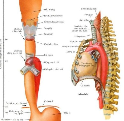
The esophagus is the first part of the digestive tube that carries food from the pharynx to the stomach, about 25cm long, relatively mobile, attached to surrounding organs by loose, flattened structures because the walls are close together. and has a tubular shape when swallowing food. The esophagus is connected to the pharynx at the level of the 6th cervical vertebrae, below the gastric outlet at the cardia, at the level of the 10th thoracic vertebra. The esophagus is located posterior to the trachea, descends to the posterior mediastinum, is located posteriorly. heart and anterior to the thoracic aorta, through the diaphragm into the abdomen, to the stomach.Anatomically, the esophagus is about 25cm long and at its narrowest point is about 1.5cm in diameter divided into 3 segments: cervical segment; thorax and abdomen. The lumen of the esophagus also has three narrow places: the junction with the pharynx at the level of the cricoid cartilage; at the level of the left aorta and left main bronchus; heart hole.
Neck segment: About 3cm long, food will run through the trachea and then to 1/3 of the neck, the food will shift to the left and down to run parallel to the trachea and then continue through the inlet behind the chest to enter the chest segment. Chest segment: About 20cm long, food entering the chest will continue through the diaphragm to enter the abdomen. Abdominal segment: About 2cm long, food has reached the abdomen, then through the foramen into the stomach.
2. What is ectopic gastric mucosa in the esophagus?
Ectopic gastric mucosa structures (ectopic gastric mucosa structures) are an area of surrounding mucosa that are salmon-pink, variable in size, and oval, round or even shaped map format. It is often observed during endoscopic evaluation of the cervical esophagus. Ectopic gastric mucosal structures may have a smooth surface or a slightly raised or concave surface with regular or irregular borders. Rarely, ectopic gastric mucosal structures may present as a convex lesion or multiple polyps. Small lesions may be covered by squamous epithelium and present without obvious changes in the upper esophageal mucosa.3. Relationship between Barrett's esophagus and ectopic gastric mucosa
Barrett's esophagus has been extensively studied for its association with adenocarcinoma and gastroesophageal reflux disease, whereas gastric mucosal structure is ectopic, a "less common relative" of enteroesophageal metaplasia, are to a large extent overlooked and their pathogenic role may be underestimated. Similar to Barrett's esophagus, ectopic gastric mucosa is often described as either a congenital condition (a vestige of the columnar mucosa of the fetal esophagus) or as an acquired condition. eg, due to an increased conversion resulting from chronic acid trauma or ruptured follicular glands. However, ectopic gastric mucosal structures, also known as gastroesophageal foreign bodies, remain a controversial topic.4. Ectopic gastric mucosa can also be found in other locations
Ectopic gastric mucosa has also been reported in other sites including the rectum and anus as well as the duodenum, jejunum, gallbladder, cystic duct, and ampulla of Vater. Their etiology and pathological features remain unexplored and unclear. In addition to the gastric mucosa, other ectopic tissues such as bronchial or pancreatic tissue can also occasionally be found in the esophagus and are frequently reported in pediatric studies, supporting the congenital hypothesis.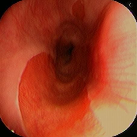
5. Esophageal gastric mucosa is a controversial disease
The proximal esophagus is rarely examined and its examination is often incomplete. Optical staining endoscopic techniques such as NBI narrowband imaging improve the detection rate of ectopic gastric mucosa, an area where their prevalence may be underestimated.Various studies have reported the correlation of these structures in the esophagus with various problems such as Barrett's esophagus, but these findings are controversial. Conflicting reports complicate the interpretation of the clinical features of ectopic gastric mucosal structures and underestimate their importance. Unfortunately, limited clinical data and statistical analysis make it difficult to draw any conclusions.
It is hypothesized that ectopic gastric mucosal lesions are associated with different esophageal and extraesophageal symptoms, and individualized diagnosis and treatment management for patients with ectopic gastric mucosa as well as in the differential diagnosis of premalignant or early cancerous lesions. Due to the lack of diagnostic capacity, there are no uniform guidelines for the management and monitoring of ectopic gastric mucosal structures. This review focuses on questions raised from the published literature on esophageal ectopic gastric mucosal structures in adults.
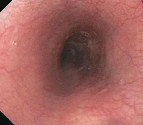
6. Ectopic gastric mucosal structure - incidental tumor or known lesion?
Currently, most endoscopists consider ectopic gastric mucosal structures in the esophagus to be a clinically unrelated entity. Others perform small studies of ectopic gastric mucosal structures or submit case reports with interesting but varied results that provide single randomization findings rather than significant correlations.6.1 Frequency of finding this structure on endoscopy The original description of an ectopic gastric mucosal structure in the upper esophagus from autopsy dates back to 1805. In this report, it was described as a unstable gastric epithelium located in the cervical esophagus.
Most cases of ectopic gastric mucosal structure consist of malformed columnar gastric mucosa usually located just below the superior esophageal sphincter or in the posterior laryngeal cartilage region of the esophagus. Similar lesions have also been found in the distal esophagus.
The incidence of ectopic gastric mucosal structures previously reported in the proximal esophagus ranged from 0.18% to 14% in endoscopic studies.
Ectopic gastric mucosal structure may not be as rare as it has been described in some previous studies because its incidence has been increased with careful bronchoscopy and careful examination of the proximal mucosa as well as by using optical endoscopy using optical staining (NBI) mode.
Furthermore, autopsies reports show significant morbidity up to 70%.
6.2 Careful exploration of the esophagus will increase the frequency of detection of this structure Therefore, this difference further demonstrates that the rate of detection of gastric mucosal structures in the esophagus during endoscopy can be high as the duration of bronchoscopy increased and when the NBI regimen was used more frequently. In most cases, the first part of the esophagus is inserted blindly, unobserved, and this area is then often neglected when the bronchoscope is removed.
>> See more: Treatment and screening for ectopic gastric mucosa in the esophagus - Article by Doctor Mai Vien Phuong - Department of Medical Examination & Internal Medicine - Vinmec Central Park International General Hospital
7. Ectopic gastric mucosa and related diseases
Ectopic gastric mucosal structures are often noticed and visualized during endoscopy in patients presenting with dyspepsia, typical or atypical symptoms of reflux disease, or a persistent burning sensation. . Other complaints related to this lesion include unexplained and persistent cough, hoarseness, chest pain, and difficulty swallowing.Furthermore, there is a variable prevalence of laryngopharyngeal reflux symptoms reported for this condition, ranging from 20% to 73.1%, which can be largely explained by sensitivity of the laryngeal mucosa to acid reflux due to ectopic gastric mucosa. Mucus secretion was also considered. Links to chronic ear or sinus infections have also been reported. In contrast, there are also authors who did not find an association between ectopic gastric mucosal structure and the presence or absence of upper esophageal symptoms in their patients. In another recent study, despite the high prevalence of ectopic gastric mucosal structures (10.9% in 239 patients), the authors determined that there was no significant association with any type. any symptoms.
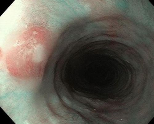
8. Clinical classification for ectopic gastric mucosa
In 2004, von Rahden et al proposed a useful clinical classification for gastric mucosal foreign bodies of the esophagusTable 1: Clinical classification of ectopic gastric mucosal structures cervical esophagus (abbreviated as CHGM) was proposed by von Rahden et al. and recommended management.
| CHGM type I | Những người không có triệu chứng với niêm mạc dạ dày lạc chỗ thực quản- trấn an bệnh nhân + theo dõi tùy chọn |
| CHGM type II không có thay đổi hình thái | Những người có triệu chứng với với niêm mạc dạ dày lạc chỗ thực quản (cảm giác nuốt nghẹn, ho, khàn giọng hoặc "biểu hiện ngoài thực quản") - trấn an và giải thích cho bệnh nhân những ngụ ý có thể xảy ra như quá mẫn thực quản + ức chế axit, prokinetic + một số trường hợp để loại trừ H Pylori nếu vẫn còn triệu chứng + đánh giá lại nội soi trong trường hợp biến chứng nghi ngờ của với niêm mạc dạ dày lạc chỗ |
| CHGM type III | Các biến chứng của với niêm mạc dạ dày lạc chỗ - liệu pháp nội soi ( ví dụ: nong bằng bóng , đốt bằng argon plasma, đốt bỏ bằng song cao tần RFA) |
| CHGM type IV | Loạn sản trong cấu trúc niêm mạc dạ dày lạc chỗ - điều trị bằng nội soi (EMR, ESD) + theo dõi |
| CHGM V | Ung thư xâm lấn trong phạm vi đầu vào- quyết định của nhóm liên ngành (bác sĩ tiêu hóa- bác sĩ ung thư- bác sĩ phẫu thuật) |
9. Classification of clinical pathology is common in which patient group?
Interestingly, several studies have reported that esophageal ectopic gastric mucosal structures are more common in men than in women. If we take into account the distribution of symptoms, for example, women are known to present more frequently with choking, this suggests that the prevalence of symptomatic ectopic gastric mucosal diseases higher in women.In the experience of the authors, it is found that there is anxiety in patients presenting with ectopic gastric mucosa mainly in women and may present as asthenia . These patients are usually classified as type 2 according to von Rahnen's classification system. The most common symptom observed by the authors was a sensation of choking without any size-dependent correlation. Similarly, other studies have found that there is a female predominance in patients with choking but conversely, concluding that it is related to reflux disease. Despite this, they recorded important information regarding size and showed that ectopic gastric mucosal structures of longer length and greater total area were more common in patients with choking. than in patients without choking.
Currently, Vinmec International General Hospital is a prestigious address trusted by many patients in performing diagnostic techniques for digestive diseases, diseases that cause chronic diarrhea, Crohn's disease, gastric mucosa Esophageal ectopic... Along with that, at Vinmec Hospital, screening for gastric cancer and gastric polyps is done through gastroscopy with Olympus CV 190 endoscope, with NBI (Narrow) function. Banding Imaging - endoscopy with narrow light frequency) results in clearer images of mucosal pathology than conventional endoscopy, detecting ulcerative colitis lesions, digestive cancer lesions In the early stages... Vinmec Hospital with modern facilities and equipment and a team of experienced experts who are always dedicated in medical examination and treatment, customers can be assured of endoscopy services. stomach, esophagus at Vinmec International General Hospital.
Please dial HOTLINE for more information or register for an appointment HERE. Download MyVinmec app to make appointments faster and to manage your bookings easily.
ReferenceRaine CH. Ectopic gastric mucosa in the upper esophagus as a cause of dysphagia. Ann Otol Rhinol Laryngol. 1983; 92 :65-66. [PubMed] [DOI] Truong LD, Stroehlein JR, McKechnie JC. Gastric heterotopia of the proximal esophagus: a report of four cases detected by endoscopy and review of literature. AmJ Gastroenterol. 1986; 81:1162-1166. [PubMed] Akbayir N , Alkim C, Erdem L, Sökmen HM, Sungun A, Başak T, Turgut S, Mungan Z. Heterotopic gastric mucosa in the cervical esophagus (inlet patch): endoscopic prevalence, histological and clinical characteristics. J Gastroenterol Hepatol. 2004; 19:891-896. [PubMed] [DOI] Borhan-Manesh F , Farnum JB. Incidence of heterotopic gastric mucosa in the upper oesophagus. Gut. 1991; 32 :968-972. [PubMed] [DOI] Tang P , McKinley MJ, Sporrer M, Kahn E. Inlet patch: prevalence, histologic type, and association with esophagitis, Barrett esophagus, and antritis. Arch Pathol Lab Med. 2004; 128 :444-447. [PubMed] [DOI] Adriana Ciocalteu, Petrica Popa, Issues and controversies in esophageal inlet patch, World J Gastroenterol. Aug 14, 2019; 25(30): 4061-4073


