This is an automatically translated article.
Posted by Specialist Doctor I Vu Thi Hanh - Radiologist - Radiology Department - Vinmec Hai Phong International General Hospital
Ultrasound is very beneficial in the diagnosis of ovarian tumors, it is a non-invasive and highly accurate imaging tool. In particular, ultrasound classification of ovarian tumors will help doctors make decisions about surgery, treatment or monitoring to ensure the best health for the patient.
1. What is the effect of ovarian ultrasound?
Ultrasound is very useful in the diagnosis of ovarian cysts. This method is non-invasive, has high accuracy, detects as well as clearly characterizes images of tumors. Ultrasound can show specific structures for each type of adnexal tumor, monitor follicular growth and tumor growth, and ultimately, thanks to those special images, the doctor The clinician may decide whether to intervene surgically or to treat medically or simply to monitor.
2. Some ways to perform ovarian ultrasound
Gynecological ultrasound can be done in two ways: Transabdominal ultrasound: usually applied to women who have not had sex, this technique requires a full bladder, so the patient must drink water. and hold urine before performing the ultrasound. However, transabdominal ultrasound images are not really clear and accurate especially in patients with thick abdominal wall or in the case of early pregnancy ultrasound or oocyte size measurement...
Ultrasound Vaginal transvaginal should be done for married or having sex women, this ultrasound has many advantages: no need to drink water first and hold urine, do it quickly and give clear and accurate images .

Hình ảnh mô tả kỹ thuật siêu âm qua đường âm đạo
3. Classification of ovarian tumors by structure on ultrasound according to IOTA:
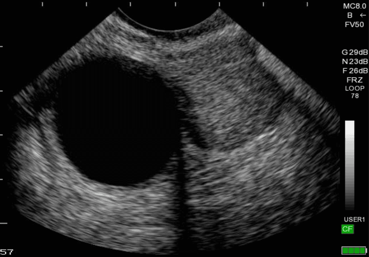
One-lobed cyst: Cyst without wall, no solid internal organization, no bud. (risk of malignancy ~ 0.6%) Polylobular cyst: Cyst with at least 1 wall. No solids, no buds (risk of malignancy ~10%). Solid multi-lobed cyst: Cyst with many lobes, walled, partially solid or with at least 1 bud. (risk of malignancy 37-43%) Solid tumor : Solid component >80%. (risk of malignancy ~ 65%) Depending on the internal composition of each tumor on ultrasound, there is a different rate of malignancy. Besides; There are some benign ovarian tumors that can be diagnosed with certainty on ultrasound such as:
Simple cyst. Endometrioid cyst. Hemorrhagic cyst. Skin cyst. Fluid retention and tubal abscesses. Pseudocyst (appears after pelvic surgery). The rule to distinguish between good and bad on ultrasound.
Homogeneous = benign. Inhomogeneity = malignancy. The rule of benign evaluation on ultrasound (5 features).
Simple one-lobed cyst. Presence of solid tissue < 7mm in greatest diameter. There is a back shadow. Smooth multilobed cyst < 100 mm. No angiogenesis.

Hình ảnh u nang buồng trứng trên kết quả siêu âm 2D
Rules for evaluating malignancy on ultrasound (5 features).
There is abdominal fluid. There are at least 4 buds. Irregular solid multilobed cyst (>100mm). Non-homogeneous solid tumor. Increased blood vessel proliferation. Ultrasound is a modern imaging tool with high accuracy to classify ovarian tumors. However, to get the most accurate ultrasound results, you need to choose a reputable performing facility, modern facilities, and a team of experienced and qualified medical professionals.
Currently, Vinmec International General Hospital systems have been using modern generations of color ultrasound machines. One of them is GE Healthcarecar's Logig E9 ultrasound machine with full options, HD resolution probes for clear images, accurate assessment of lesions. In addition, a team of experienced doctors and nurses will greatly assist in the diagnosis and early detection of abnormal signs of the body in order to provide timely treatment.
Please dial HOTLINE for more information or register for an appointment HERE. Download MyVinmec app to make appointments faster and to manage your bookings easily.


