This is an automatically translated article.
The article was written by MSc Nguyen Thuc Vy - Department of Diagnostic Imaging - Vinmec Nha Trang International General HospitalComputed tomography coronary angiography with intravenous contrast coronary angiography (CTA) provides information on coronary artery anatomy and the extent of stenosis/occlusion.
1. Definition
However, to decide whether coronary artery stenosis affects hemodynamics (blood volume to the myocardium after stenosis) and whether reperfusion intervention is needed, cardiologists still need other methods to Assessments such as:
Invasive methods: FFR (coronary artery reserve fraction) is called the gold standard for hemodynamic assessment after stenosis, performed during the time of interventional angiography .
Non-invasive method :
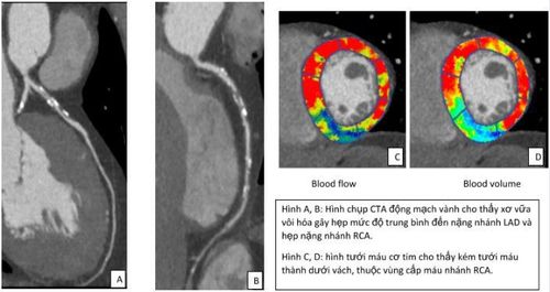
Stress echocardiography SPECT/ PET MRI myocardial perfusion CT myocardial perfusion CT coronary artery - myocardial perfusion is the method to decide whether coronary artery stenosis is reduced blood flow to the myocardium without sending the patient to another clinic, on a different day, and performing other diagnostic methods. This technique will provide the doctor with information about the myocardial perfusion level with color coding of the hypoperfusion area, making it easier for the doctor to assess the degree of myocardial hypoperfusion, thereby deciding whether to intervene. coronary revascularization or not.

2. Implementation process
There are many techniques to assess myocardial perfusion, currently at Vinmec Nha Trang, we are using dynamic myocardial perfusion techniques using drugs to create stress dynamic myocardial perfusion.
With this technique, the patient will be placed two lines in both hands, one line for high-speed contrast injection and one line for adenosine infusion. The patient will be injected with contrast twice:
+ 1st time: coronary computed tomography to evaluate the anatomy as well as the degree of coronary stenosis/occlusion
+ 2nd time: the patient will receive adenosine infusion intravenously for 3 minutes. Adenosine is a vasodilator, increasing blood flow to the coronary arteries and myocardium, and the administration of adenosine exposes areas of myocardial hypoperfusion similar to that of an exercise patient. During the infusion of the drug, the patient will feel a rapid heartbeat and palpitations similar to when exercising. Patients will be monitored by doctors for symptoms, pulse, ECG and blood pressure during adenosine infusion. After 3 minutes of infusion of adenosine, the patient will receive a second contrast injection to capture myocardial perfusion.
Two injections of contrast medium about 15-20 minutes apart to allow the contrast from the first injection to be completely cleared from the heart.
Prepare the patient before the scan:
Stop preparations containing xanthine (tea, coffee), chocolate, theophylline, dipyridamole 24-36 hours before the scan. Discontinue ViagraTM, CialisTM or LevitraTM, 24 to 72 hours before the scan. Having symptoms of chest pain suspected of acute coronary syndrome should be tested for CK, CKMB, troponin. Preoperative electrocardiogram (ECG) Indications:
Stable angina Patients at risk for coronary artery disease or atypical chest pain Coronary risk stratification Before surgery with or without cardiac involvement After coronary intervention or revascularization procedure

Contraindications:
In the following cases, it will not be possible to perform an exercise myocardial perfusion technique:
The patient has symptoms of unstable angina or acute chest pain suspected of developing acute coronary syndrome . Patients with contraindications to contrast media and adenosine. In the case of patients with contraindications to nitroglycerin (used to dilate coronary arteries before coronary computed tomography), nitroglycerin should not be used during the scan. The patient will be examined by a cardiologist to evaluate the cardiovascular symptoms, if there are no above contraindications and coronary artery disease is suspected, the patient will be appointed by the doctor to perform the irrigation technique. myocardial blood flow on exertion.
Comparison of CT myocardial perfusion technique with other methods:
CT myocardial perfusion has similar sensitivity and specificity to myocardial perfusion scintigraphy (SPECT).
The level of radiation exposure is comparable to SPECT and can be lower using radiation dose reduction techniques in newer CT machines.
Sensitivity and specificity are close to the threshold of myocardial perfusion MRI.
CT myocardial perfusion is a modern technique that has been used in many countries around the world. Combined coronary CTA and myocardial perfusion is the only method that provides information on coronary artery stenosis and hemodynamics in a single survey.
Vinmec Nha Trang has been implementing this technique with the 512-slice CT GE Revolution machine, a modern machine with 16 cm coverage in one scan, helping to survey the entire myocardium in one rotation. bring reliable results to support the diagnosis and treatment of patients with coronary artery disease.
Please dial HOTLINE for more information or register for an appointment HERE. Download MyVinmec app to make appointments faster and to manage your bookings easily.


