This is an automatically translated article.
The article is professionally consulted by Internal Cardiologists and Interventional Cardiologists - Cardiovascular Center - Vinmec Central Park International General Hospital.An arrhythmia is an abnormal condition of the heart rhythm. Arrhythmia occurs in many different pathological contexts, diagnosis and classification of arrhythmias in clinical practice are mainly based on electrocardiogram.
1. What is an arrhythmia?
Heart is one of the essential organs in the human body. The heart's contractions are rhythmic and regular thanks to the impulse conduction system and the integrity of the heart muscle cells. Cardiac muscle cells are a special type of cell that mediates both smooth and skeletal muscle cell types. Between the cardiac muscle cells there are junctions, similar in structure to the syncytial cells. Thanks to this feature, when a stimulus occurs, the rate of propagation occurs faster between cardiac muscle cells.
The heart's electrical impulse conduction system has the ability to spontaneously beat and conduct electrical impulses through the chambers of the heart to help the heart muscle fibers contract synchronously, ensuring the bioelectrical activity of the heart. Components of the conduction system include:
● Sinus node : located in the right atrium muscle. This is the heart's master node, capable of spontaneously emitting a pulse with a frequency of about 80-100 times / minute. The sinus node is under the control of the sympathetic and parasympathetic nervous systems.
● Atrioventricular node: located to the right of the lower part of the atrial septum, capable of generating pulses with a slower frequency than the sinus node, about 40-60 times/minute.
● Bundle of His: runs along the interventricular septum, connecting from the atrioventricular node to the ventricles. The bundle of His runs in the interventricular septum for a short distance of about 1 centimeter, then divides into left and right branches. These two branches run down the right and left ventricles, respectively, giving rise to many small branches that weave between the ventricular muscle fibers called Purkinje fibers. The bundle of His, its two right and left branches, and the Purkinje fibers are also capable of pulsating but with a very low frequency, about 20-40 beats/min.
Cardiac arrhythmia is a medical condition that occurs due to a disturbance in the bioelectrical activity of the heart. When sinus node pacing and/or the activity of the impulse conduction fibers are abnormal, arrhythmias occur.

Các dạng rối loạn nhịp tim rất đa dạng
2. Causes of arrhythmia
When a patient comes to a medical facility because of an arrhythmia, the doctor needs to ask for a history, do a thorough physical examination and order many necessary tests because the causes of arrhythmia are very diverse. Some common diseases can cause cardiac arrhythmias such as:
● Infections: some infectious diseases affect the contractile activity and conduction of the heart's impulses, the most common being rheumatic fever. Diphtheria , typhoid , ... are also other infectious diseases that are thought to cause cardiac arrhythmias
● Intoxication : antiarrhythmic drugs are used improperly, leading to a state of arrhythmia. Poisoning can become counterproductive and cause arrhythmias such as beta blockers, digitalis, procainamide, reserpine, ...
● Electrolyte disturbances: hyperkalemia, hypocalcemia, ...
● Global pathology body: diabetes, hyperthyroidism, ...
● Cardiomyopathy: myocardial injury, myocardial infarction, congenital heart diseases such as tetralogy of fallot, patent ductus arteriosus, ...
● Disorders autonomic nervous system
Postoperative sequelae related to thoracic surgery
● Genetic factors.
Nhiễm khuẩn có thể gây nên rối loạn nhịp tim
3. Classification of arrhythmias
The clinical forms of arrhythmias are very diverse. There are many ways to classify arrhythmias, based on the location of the abnormal rhythm or the heart rate. Distinguishing types of arrhythmias according to heart rate is an easy initial approach, including:
Arrhythmias with normal and regular rates: First degree atrioventricular block, accelerated autoventricular rhythm, ventricular splicing, supraventricular tachycardia with conduction block, atrial extrasystoles, atrial flutter associated with atrioventricular block, aberrant conduction, drug toxicity Arrhythmias with normal and irregular rates: Second degree atrioventricular block, irregular conduction supraventricular tachycardia such as atrial fibrillation, atrial flutter, sinus arrhythmias with or without conduction block or aberration, drug toxicity, hyperkalemia Arrhythmia with slow and regular heart rate: sinus bradycardia, autoventricular rate, junctional bradycardia, complete atrioventricular block with or without junctional escape rhythm, supraventricular tachycardia with severe atrioventricular block arrhythmia with slow and irregular frequency: sinoatrial block, second-degree AV block, group 1 or 2, bradycardia, arrhythmias with a rapid and regular rate: tachycardia ang, ventricular tachycardia, sinus node re-entry tachycardia, aberrant conduction tachycardia, drug toxicity, hyperkalemia Arrhythmia with rapid and irregular rate: supraventricular tachycardia, extrasystolic tachycardia atrial and ventricular, ventricular fibrillation, ventricular tachycardia, multifocal atrial tachycardia, atrial fibrillation and atrial flutter with accessory conduction pathways, ...
4. Electrocardiogram characteristics of common types of arrhythmias
Electrocardiogram is a means of recording the electrical activity of the heart, helping to diagnose and classify different types of arrhythmias.
4.1 Sinus tachycardia
The electrocardiogram of sinus tachycardia still has all the features of a sinus rhythm such as the presence of a normal P wave, with a complete QRS complex. Heart rate calculated on paper recorded over 100 beats/min, steady.
4.2 Sinus bradycardia
Patients with sinus bradycardia are often admitted to the hospital with clinical symptoms such as syncope, feeling dizzy, and dull. The electrocardiogram electrodes were fitted with all the features of a sinus rhythm, such as P wave, PR interval and QRS complex with normal shape, combined with bradycardia below 60 beats/min.
4.3 Atrial fibrillation
When atrial fibrillation occurs, on the electrocardiogram no longer shows the normal P wave image, instead there are small fluttering f waves, with a rapid rate of about 300-600 beats/min. The QRS complex varies in irregular amplitude.
4.4 Crazy ears
Similar to atrial fibrillation, atrial flutter is a type of arrhythmia, belonging to the subgroup of supraventricular tachycardias. The electrocardiogram of atrial flutter no longer shows normal P waves, instead there are F waves with sawtooth shape, lower frequency than atrial fibrillation, about 200-300 beats/min.
4.5 Paroxysmal supraventricular tachycardia
The heart rate observed on the electrocardiogram is about 180 beats/min. The P wave is no longer visible because it is mixed with the normal, regular QRS complex. The ST segment may change in the direction of depression.
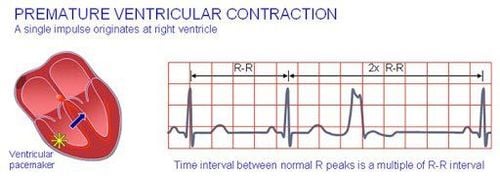
4.6 Ventricular tachycardia
Electrocardiogram of ventricular tachycardia only observed dilated ventricular complexes, the frequency is about 120-160 beats/min. The atrial rhythm is slow and separate from the ventricular rate, making it difficult to observe the P wave.
4.7 Ventricular fibrillation
This is an emergency arrhythmia because it can easily lead to sudden death. The electrocardiogram of ventricular fibrillation has a polymorphic ventricular complex disorder, without the normal shape of the QRS. In this case the heart no longer contracts normally, but only vibrates.
Vinmec International General Hospital has applied electrocardiogram technique in examination and diagnosis of many cardiovascular diseases. Electrocardiogram at Vinmec is carried out methodically and in accordance with standard procedures by a team of highly qualified medical professionals, modern machinery system, thus giving accurate results, making a significant contribution to the identification. disease and disease stage.
Please dial HOTLINE for more information or register for an appointment HERE. Download MyVinmec app to make appointments faster and to manage your bookings easily.
Reference article source: Vietnam Cardiology Association