This is an automatically translated article.
The article is professionally consulted by Master, Doctor Luu Thi Bich Ngoc - Department of Diagnostic Imaging and Nuclear Medicine - Vinmec Times City International Hospital
Spine infection is a disease that causes serious damage to the spine and negatively affects the patient. Therefore, early detection and treatment can prevent damage. Many patients wonder if "can a spinal infection be diagnosed with an MRI scan?".
1. Learn about spinal infections
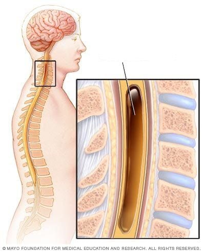
The spine is the pillar bone, which plays an important role in the body. The spine is composed of vertebrae. Each vertebra has 3 parts, including: vertebral body, vertebral arch and vertebral foramen. Between the two vertebral bodies there is a spinal disc.
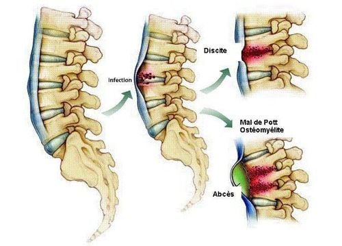
Infection of the spine is a common pathology in bone infections. Pathology usually originates from distant foci of infection, then hematogenously spreads to the spine via the arterioles.
Causes of spinal infection such as:
Spine infection caused by pyogenic bacteria: This condition is often found in the lumbar spine or a segment of the spine. Tuberculosis infections: Most common in the chest area and less commonly in the lumbar region. Even with imaging, it is difficult to distinguish. Brucella spondylitis: Brucella infection is an infection in animals with worldwide distribution. In humans, brucellosis is commonly acquired from contaminated animal products by hand or by consuming unpasteurized dairy products. The spine is the most susceptible site for brucellosis in the musculoskeletal system. Aspergillus spondylitis: Aspergillus is a saprophytic fungus that can cause spinal infections in immunocompromised patients. It is also rare in immunocompromised individuals.
2. What is MRI technique?
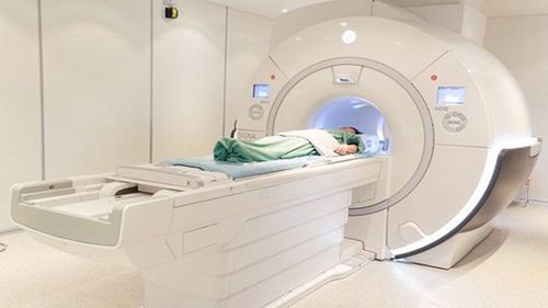
MRI is also known as Magnetic Resonance Imaging. This is an imaging technique that does not use X-rays, but uses a magnetic field and radio waves. Therefore, it is very safe for the patient, with no risks. MRI is a modern method widely applied in the world, bringing detailed and multidimensional diagnostic value.
However, some metal devices implanted in the human body may be affected by strong magnetic fields. This method is not indicated for women who are in the first 12 weeks of pregnancy.
3. Can a spinal infection be diagnosed with an MRI scan?
According to experts, spinal infection can be diagnosed with magnetic resonance imaging. Besides, this technique also plays a role in assessing disease status, distinguishing infection and other diseases. Because the MRI technique has high sensitivity and specificity. According to experts, the specificity of MRI also depends on the signal as well as the anatomical distribution of the infection.
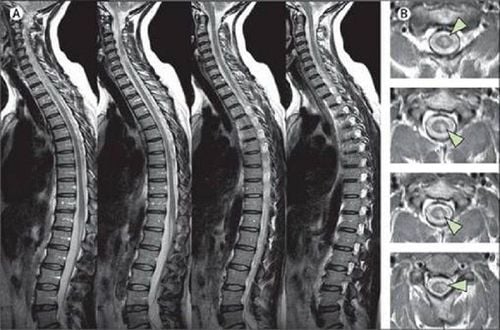
Spinal infection is diagnosed on MRI when the vertebral body surface shows signs of destruction, damage to two consecutive vertebrae and discs.
Infection of the spine is a complex disease that shares many features with non-infectious or degenerative inflammation. Therefore, it is difficult to accurately diagnose the disease. Specialists need to know in detail the atypical MRI signs of spinal infections to avoid misdiagnosis and ineffective treatment regimens.
Here are some signs of spinal infection on MRI images:
MRI images show bacterial spondylitis To evaluate for bacterial spondylitis, your doctor will order an X-ray Magnetic Resonance. The imaging results obtained from this method allow the evaluation of epidural inflammation and spinal cord compression. At the same time, the dural sheath image is also clearly displayed.
MRI will help doctors monitor the patient's response to treatment and make clinical decisions. Specifically, MRI imaging will monitor soft tissue remission and bone marrow fat deposition. This is a sign that the patient's condition has improved clinically.
MRI image shows tuberculous spondylitis To distinguish tuberculous spondylitis from pyogenic bacteria, the doctor will order an MRI scan. If the images obtained have the following signs, it will result in tuberculous spondylitis: smooth, thin abscess wall, presenting under the three or more vertebral ligaments; The patient suffered damage to many vertebrae and the whole vertebrae. In addition, MRI also allows to observe some abscesses near the spine.
MRI image showing brucellosis MR features characteristic of brucellosis include low lumbar spine predominance, intact vertebral structure despite evidence of diffuse spinal osteomyelitis, markedly increased disc signal on T2-weighted images and post-injection images, damage to the intertemporal joint.
MRI images show spondyloarthritis caused by aspergillus According to experts, spondylitis caused by aspergillus will have the same imaging results as spondyloarthritis caused by pyogenic bacteria. However, the disc in an aspergillus infection will not become inflamed or invasive. In addition, there is inflammation caused by aspergillus with manifestations such as: non-intense disc on T2W and STIR; The disk kernel slot can be preserved.
Atypical imaging patterns of spinal infections Recognizing the atypical imaging patterns of spinal infections is important in the appropriate clinical setting to avoid delayed diagnosis. Atypical patterns include involvement of only one vertebral body, one vertebral body and one disc, and two vertebral bodies without disc involvement.
To protect their own health, patients need to go to a reputable hospital to conduct examination and treatment as soon as there are signs of spinal infection.
Currently, Vinmec International General Hospital is one of the leading prestigious hospitals in the country, trusted by a large number of patients for medical examination and treatment. Not only the physical system, modern equipment: 6 ultrasound rooms, 4 DR X-ray rooms (1 full-axis machine, 1 light machine, 1 general machine and 1 mammography machine) , 2 DR portable X-ray machines, 2 multi-row CT scanner rooms (1 128 rows and 1 16 arrays), 2 Magnetic resonance imaging rooms (1 3 Tesla and 1 1.5 Tesla), 1 room for 2 levels of interventional angiography and 1 room to measure bone mineral density.... Vinmec is also the place to gather a team of experienced doctors and nurses who will greatly assist in diagnosis and detection. early signs of abnormality in the patient's body. In particular, with a space designed according to 5-star hotel standards, Vinmec ensures to bring the patient the most comfort, friendliness and peace of mind.
Please dial HOTLINE for more information or register for an appointment HERE. Download MyVinmec app to make appointments faster and to manage your bookings easily.


