This is an automatically translated article.
This article is professionally consulted by Master, Doctor Luu Thi Bich Ngoc - Radiologist - Department of Diagnostic Imaging and Nuclear Medicine - Vinmec Times City International Hospital.A condition in the brain in which clumps of tissue are produced by abnormally fast-growing cells called brain tumors. Brain tumors can be benign or they can be malignant. In fact, brain tumors are often detected late, so they face many difficulties in management, patients often have a poor prognosis and leave quite serious consequences.
1. Brain Tumor Overview
There are many different types of brain tumors, of which less than 50% of intracranial tumors are intracranial tumors, the rest are tumors originating from the meninges, cranial nerves, pituitary gland, metastatic tumors... By removing water that brain tissue contains more than other tissues, tumors in the skull, especially tumors of brain tissue often have slow progression, the brain will have time; Clinical symptoms will also appear gradually and insidious. If in the case of a brain tumor with infiltrative and rapid growth, the clinical picture will be more severe and clear. This type of brain tumor often compresses the surrounding circulatory system, causing cerebral edema. due to the vascular mechanism.In addition, because of the existing or sudden increase in intracranial pressure syndrome, some types of tumors have fluid cysts inside, the blood vessels proliferate much, easily causing bleeding inside the tumor, causing The course of the disease resembles an acute illness. In fact, the skull has a closed volume divided into many compartments, separated by walls such as the crescent of the brain, the tent of the cerebellum; The tumor will appear when the pressure in the compartments is not equal, resulting in the brain tissue being pushed from one cavity to the other as in the case of brain tissue in the hemisphere of the brain with the tumor being pushed through under the crescent of the brain. on the opposite side, the temporal lobe hippocampus passes through the free border of the cerebellar tent, the cerebellar amygdala enters the occipital foramen for pressure equalization; These are forms of "brain loss" with very serious consequences, sometimes leading to death.
Brain tumors often present 3 types of symptoms, including:
General signs: Typical signs are seizures Increased intracranial pressure Symptoms are due to the histology of the mass and the location of the tumor About 40% of brain tumors present with general symptoms such as seizures and increased intracranial pressure. The first sign of the common symptom is a seizure. This is a symptom that immediately suggests a brain tumor, but in fact, at present, in our country, signs of this disease have not been detected much.
A set of signs indicating the presence of a tumor in the skull causing a diffuse headache, which becomes more intense and continuous later on, the patient can vomit easily from waking up , suffers from papilledema, a sluggish mental state known as intracranial hypertension syndrome. Signs such as finger prints on conventional cranial X-rays; on the radiographs, it is possible to see the back of the pituitary with wear and tear, craniofacial defects or hyperplasia of the skull; Abnormal calcifications and calcifications of the pineal gland deviating from its normal position are laboratory findings that may suggest a brain tumor. In our country, these are valuable signs in the diagnosis of brain tumors, but in other countries, calcification is less common.
Disease symptoms related to histological properties of tumors also have certain differences. Tumor infiltrates cause the disease to progress quite quickly, and at the same time cause hemodynamic disturbances, creating cerebral edema in malignant tumors with a short history such as neuroblastoma. In contrast, the disease usually progresses very slowly over many years, the tumor has a lot of vascular proliferation for benign tumors, especially meningioma. In our country, many cases of tumors can reach unexpected large sizes because most of the patients are detected and diagnosed late. Two types of brain tumors, malignant and benign, usually occur in patients in adulthood, a few occur in middle age. Most tumors develop in the posterior fossa in preschool children. Craniopharyngeal tumors are usually congenital tumours, so they are more common in children.
Symptoms have different manifestations due to the location of the tumor. Because this lobe is larger than the other lobes, tumors of the frontal lobe are relatively common. In fact, it is possible to direct a diagnosis of a local brain tumor by the synthesis of focal symptoms, which means that the tumor's location in the skull and in the brain tissue can be determined based on it.

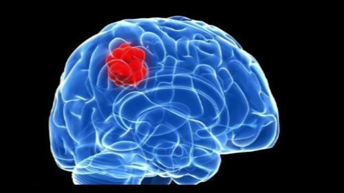
Dấu hiệu điển hình của u não là những cơn động kinh
2. Detecting brain tumors through X-rays
2.1 What is an X-ray? X-ray is a familiar term for many people. High-energy radiation is emitted from X-rays, X-ray machines emit X-rays - these rays can penetrate body surfaces, soft tissues and even body fluids.X-rays produce black and white images of organs such as lungs, heart, bones, blood vessels.. after passing through so many layers of the human body. The doctor will rely on the images taken by the X-ray machine to find abnormalities in these parts, detect diseases and plan treatment for the patient. X-rays can't penetrate dense tissue because they interfere with X-rays.
To examine the bones of the skull, including the bones of the face, nose, and sinuses, your doctor will examine the images obtained from the scan. X-ray. This is considered an easy, fast and effective way to help your doctor examine the area that contains your most important organ - your brain. As for the case of brain tumor, X-ray is indicated to evaluate the invasion and destruction of the brain tumor into the bone.
Cranial X-ray is often used in cases of suspected head trauma, showing damaged skull and facial bones.
X-ray is one of the steps that can be indicated to diagnose diseases, especially bone and joint diseases. X-rays can help us detect lesions that cannot be seen with the naked eye.
2.2. Diagnosis of brain tumor through X-ray The doctor will order an X-ray of the skull in cases of falls, strong head impacts or trauma caused by traffic accidents. The images will show if the skull is fractured or broken. Based on that, the doctor will make a more reasonable treatment plan.
In particular, a head X-ray can help diagnose brain tumors. X-rays help doctors detect abnormal changes in the skull bones caused by tumors, detect calcium deposits in brain tumors (bone resorption, bone thickening due to metastasis or invasion). of brain tumors).
Besides recorded clinical symptoms and X-ray methods, scientists can use other means to diagnose brain tumors such as: Brain ultrasound, brain scan recording, electroencephalogram recording , computed tomography, magnetic resonance imaging of the nuclei, brain scan by positron emission, magnetic field recording of the brain or cranial MRI....At present, the diagnosis of brain tumors has become more popular. made easier and more precise thanks to the achievements of science and medicine, gradually replacing the old means and techniques.

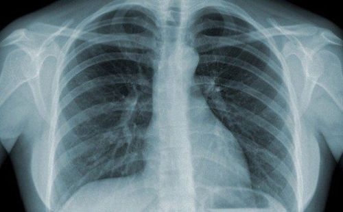
Chụp X- quang có thể phát hiện những bất thường trong não và chẩn đoán u não
3. Does head X-ray affect health?
Head X-ray technique is very commonly used in medicine today, especially in the diagnosis of bones and joints. So whether your health will be affected when conducting a head X-ray is the question of many people today.For health, X-ray method does not affect anything, but X-ray is very toxic and dangerous. X-rays have high radiation energy, which can cause damage to our health. X-rays can affect fertility. If X-rays are overused, sensitive parts such as genitals, bone marrow, skin... can be affected the most.
You can have hair loss, skin burns, the most undesirable case is cancer if X-rays are taken continuously for a short period of time. However, an X-ray won't do any harm if you know the time intervals. Head X-rays in particular and other types of X-rays in general should only be taken when prescribed by a doctor.
Because of the possible effects on the fetus, pregnant women should avoid X-rays. In cases where X-rays are required, it is necessary to follow the instructions of the doctor or use modern equipment.
Doctors need to be more careful with children patients. You should talk to your doctor about your child's condition and the risks that radiation may pose to your child.
To ensure that you absorb the minimum amount of X-ray radiation during your head X-ray, it is important to follow safe procedures and conditions.
However, exposure levels that are considered safe for adults may not be safe for the developing fetus. Talk to your doctor about this if you are pregnant or trying to get pregnant.
Currently, X-ray is an imaging method performed routinely at Vinmec International General Hospital, including head X-ray technique. The X-ray procedure at Vinmec is carried out methodically under the guidance of the medical team of the Department of Diagnostic Imaging. In addition, Vinmec is now equipped with the most modern X-ray machine, meeting international standards for true and clear images, helping doctors to accurately diagnose the disease and the stage of the disease, from It has an effective treatment direction, creating a sense of safety for the patient.
Any questions that need to be answered by a specialist doctor as well as customers wishing to be examined and treated at Vinmec International General Hospital, please register for an online examination on the Website for the best service.
Please dial HOTLINE for more information or register for an appointment HERE. Download MyVinmec app to make appointments faster and to manage your bookings easily.