This is an automatically translated article.
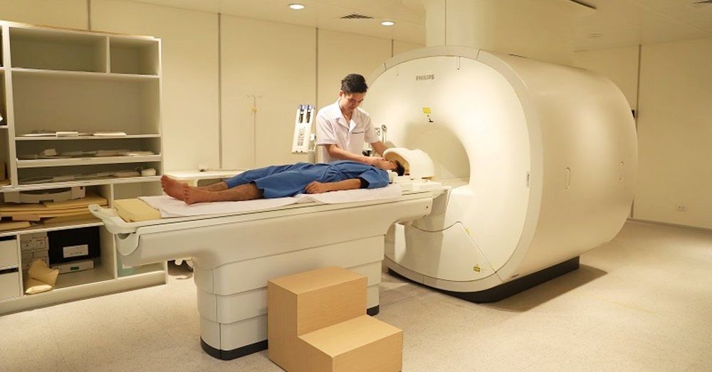
Through the symptoms of knee pain, swelling of the knee joint, difficulty in walking, ... to further confirm the diagnosis of patellar tendon rupture, the doctor will assign the patient to take a routine X-ray film. or magnetic resonance imaging (MRI) of the knee joint.
1. Causes of patellar tendon rupture
The patellar tendon is a tendon that attaches from the lower pole of the kneecap to the top of the tibia. Also attached to the pole above the kneecap is the quadriceps tendon. Therefore, the quadriceps tendon and patellar tendon have a role like a pulley, when the quadriceps muscle contracts, the patellar tendon is stretched, making it easy for people to stretch the knee.
However, patellar tendon can also be ruptured, the cause of patellar tendon rupture is usually due to direct trauma to the knee or sudden knee flexion during sports or daily activities. In addition to traumatic causes, some musculoskeletal conditions can cause the patellar tendon to lose its strength, leading to rupture. Some of the causes are:
Patellar tendinitis : Patellar tendonitis is mainly caused by repetitive movements or can be caused by corticosteroid injections directly or around the patellar tendon, which increases the risk of tearing the patellar tendon. higher tea. Chronic diseases: Chronic diseases such as chronic kidney failure, rheumatoid arthritis, diabetes, infections, metabolic diseases can lead to poor blood supply, thereby weakening the patellar tendon. and break easily. Surgery around the knee: In some cases, previous surgery around the knee (knee replacement, cruciate ligament reconstruction) can cause patellar tendon rupture. Most of the time, after breaking the patellar tendon, the patient is often subjective and thinks that the injury is small, after a few days it will heal, which makes the injury worse and worse, affecting daily activities and movement. daily.

Kỹ thuật chụp MRI là phương pháp chẩn đoán đứt gân bánh chè hữu ích
2. What medical technique is a patellar tendon rupture diagnosed?
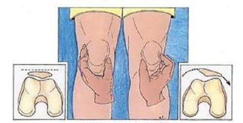
After the patellar tendon ruptures, the patient will feel knee pain, the knee joint also gradually swells. Accompanied by symptoms such as palpating the concave area at the lower pole of the kneecap, the knee is also bruised, often cramping, difficult to fully extend the knee, difficulty walking, and notice that the kneecap is higher than before, so I went to a medical facility for examination.
Through the above clinical symptoms, in order to diagnose patellar tendon rupture most accurately, the doctor will assign the patient a routine X-ray or magnetic resonance imaging (MRI) of the knee joint. Because these two imaging techniques show:
On routine radiographs with the lateral position, the patella is in a higher position than normal. If in the case of complete rupture of the patellar tendon, only routine radiographs can accurately diagnose the injury; MRI technique is a useful method of diagnosing patellar tendon rupture, because this technique can display more detailed and clearer images of soft tissue injuries, determine the extent and location of the tendon. broken tea. In some cases, an MRI can also help the physician differentiate between lesions with similar clinical symptoms.
Is a common injury in daily life and work, but due to the patient's subjectivity and late examination, it causes difficulties in treatment. Therefore, when there is an injury in the knee area with difficulty in stretching the knee after being treated with anti-inflammatory, anti-edematous and pain-relieving drugs... but the symptoms are still not relieved, it is necessary to immediately seek medical attention. specialists for timely examination and treatment, avoiding dangerous complications. Currently, Vinmec International General Hospital is one of the leading prestigious hospitals in the country, trusted by a large number of patients for medical examination and treatment. Not only the physical system, modern equipment: 6 ultrasound rooms, 4 DR X-ray rooms (1 full-axis machine, 1 light machine, 1 general machine and 1 mammography machine) , 2 DR portable X-ray machines, 2 multi-row CT scanner rooms (1 128 rows and 1 16 arrays), 2 Magnetic resonance imaging rooms (1 3 Tesla and 1 1.5 Tesla), 1 room for 2 levels of interventional angiography and 1 room to measure bone mineral density.... Vinmec is also the place to gather a team of experienced doctors and nurses who will greatly assist in diagnosis and detection. early signs of abnormality in the patient's body. In particular, with a space designed according to 5-star hotel standards, Vinmec ensures to bring the patient the most comfort, friendliness and peace of mind.
Please dial HOTLINE for more information or register for an appointment HERE. Download MyVinmec app to make appointments faster and to manage your bookings easily.


