This is an automatically translated article.
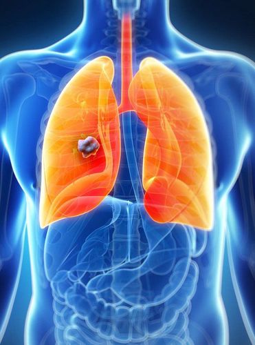
Bronchiectasis is a chronic, irreversible dilatation of one or more bronchi. Bronchiectasis is diagnosed by chest X-ray and CT scan of the chest.
1. What is bronchiectasis?
Bronchiectasis is a pathological condition of the bronchi, due to the destruction of the structures of the bronchial wall or weakening and dilation. The damage of chronic bronchiectasis is irreversible damage, unlike acute bronchiectasis, which is temporarily reversible in some acute infections such as viral infections, bronchiolitis, and bronchiolitis. Bronchiectasis ...
Bronchiectasis is divided into: saccular bronchiectasis, cylindrical bronchiectasis and rosary bronchiectasis.
Bronchiectasis can be congenital, inherited or acquired. Bronchiectasis can be focal and limited to one or one lobe of the lung, or it can extend to multiple lobes in one or both lungs.
2. Diagnosis of bronchiectasis
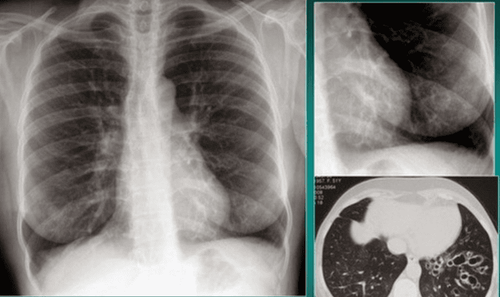
Hình ảnh giãn phế quản khu trú LIG
On standard chest radiograph: signs suggestive or confirmed for diagnosis of bronchiectasis may be seen in some cases of severe and severe bronchiectasis. However, the definitive diagnosis of bronchiectasis usually has to be based on 1mm class computed tomography, high resolution images of bronchiectasis on X-ray:
The bronchial walls form parallel lines (rails). The volume of the lobe of the lung with bronchiectasis is reduced, and the vascular opacities of the lung are fused together if there is atelectasis. There are small light spots like honeycombs, there may be bright spots with horizontal water level usually no more than 2 cm. Image of recurrent pneumonia in the cold season around the bronchiectasis area. Tubular opacities represent bronchi filled with mucus, pus. About 7 - 30% of cases with a standard chest x-ray show nothing abnormal. High-resolution, thin-layer computed tomography is the gold standard in the definitive diagnosis of bronchiectasis. Possible signs: The inner diameter of the bronchus is larger than the accompanying artery. Non-descending bronchi are prescribed when the upper bronchus is 2 cm long with a diameter similar to that of the bronchus that have divided into that bronchus. The bronchus is seen less than 1 cm from the pleura of the chest wall. The bronchus is seen close to the mediastinal pleura.
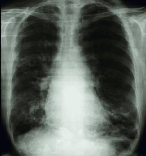
Hình ảnh giãn phế quản trên X quang
Vinmec International General Hospital is a high-quality medical facility in Vietnam with a team of highly qualified medical professionals, well-trained, domestic and foreign, and experienced.
A system of modern and advanced medical equipment, possessing many of the best machines in the world, helping to detect many difficult and dangerous diseases in a short time, supporting the diagnosis and treatment of doctors the most effective. The hospital space is designed according to 5-star hotel standards, giving patients comfort, friendliness and peace of mind.
To register for an examination at Vinmec International General Hospital, you can contact the nationwide Vinmec Health System Hotline, or register online HERE.


