This is an automatically translated article.
This article was written by Specialist Doctor II Khong Tien Dat, Doctor of Radiology - Department of Diagnostic Imaging - Vinmec Ha Long International Hospital.Brain perfusion magnetic resonance imaging is increasingly indicated because of its safety and ability to reproduce sharp images. The purpose of cerebral perfusion magnetic resonance imaging includes assessment of cerebral perfusion status and function of each area of brain parenchyma. Cerebral perfusion magnetic resonance imaging is indicated primarily in the setting of acute ischemic stroke and pathological brain tumors.
1. Purpose of cerebral perfusion magnetic resonance imaging
Magnetic resonance imaging is increasingly asserting its role and position with the ability to bring detailed images of high quality. Brain perfusion magnetic resonance imaging or cerebral perfusion MRI is a relatively new technique that provides information on cerebral hemodynamic parameters such as relative cerebral blood volume (rCBV) and cerebral blood flow (CBF). Surveying and evaluating the function of the cerebrovascular system, as well as evaluating the brain parenchyma function is the purpose of cerebral perfusion magnetic resonance imaging. Cerebral perfusion MRI can be performed with or without contrast.Cerebral perfusion magnetic resonance imaging is evaluated as a safe imaging modality. Patients are not exposed to X-rays as when computed tomography, so it can be applied to sensitive subjects such as pregnant women and children. Contrast used in perfusion MRI is less likely to cause allergic reactions or drug shock when compared with those used in computed tomography.

Chụp cộng hưởng từ ngày càng khẳng định vai trò và vị trí của mình
2. Indications/contraindications for cerebral perfusion MRI
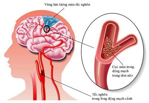
Brain perfusion MRI is the preferred choice when you want to evaluate the circulation status of the brain and the function of the brain parenchyma at the time of the survey. Therefore, the indications for cerebral perfusion magnetic resonance imaging are often related to cerebrovascular diseases or lesions affecting the vascular system. Together with other imaging diagnostic tools such as transcranial ultrasound, computed tomography, cerebral perfusion MRI, it aids in the diagnosis and monitoring of treatment results in the following pathologies:Brain tumor Ischemic stroke: In the acute phase of cerebral infarction, the purpose of cerebral perfusion magnetic resonance imaging is to evaluate the ischemic status of each brain parenchyma area. In which, it is necessary to detect brain regions that are not really dead and have the ability to recover to have a more appropriate treatment strategy. Cerebrovascular malformations Despite its many advantages over other imaging modalities, cerebral perfusion MRI is not routinely indicated for all patients because of its unique risks. . Contraindications of cerebral perfusion MRI that should be noted include:
People carrying artificial replacement materials made of metal or magnetic inside the body such as artificial heart valves, pacemakers, snails artificial ears, screws or splints used to fuse bones, hemostatic clips in cerebral aneurysms ... Nowadays many manufacturers have added information about contraindication to MRI in their manuals. surname. Patients need to ask and understand information to easily coordinate with medical staff. People with claustrophobia or fear of the dark find it difficult to cooperate when conducting magnetic resonance imaging because they have to lie down for a long time in one position under the imaging chamber. This condition can be alleviated by the use of sedatives. The person has a metal foreign body in the eye sockets or head area. People who are suffering from serious acute diseases need to be resuscitated. In order to ensure patient safety and minimize image disturbances, medical staff and patients need to cooperate in examining before taking pictures, taking medical history and medical history. used carefully to rule out contraindications.
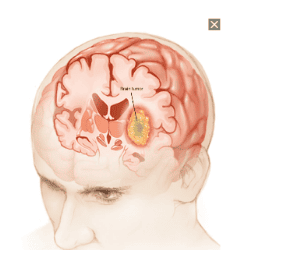
Chụp MRI tưới máu não giúp chẩn đoán u não
3. What do patients need to prepare before taking MRI of cerebral perfusion?
Before taking magnetic resonance imaging in general or brain perfusion MRI in particular, patients need to follow the principles that medical staff have advised to keep themselves safe and coordinate to have good image quality. . Things to note include:Persons undergoing cerebral perfusion magnetic resonance imaging do not need to fast before. In case the doctor orders an MRI with contrast, the patient needs to remember not to eat or drink at least 4 hours before the time of the scan. Patients need to be consulted and explained in detail, so ask for information about the technique they are made. Cooperate in exploiting relevant history information to rule out contraindications to perfusion MRI. Prepare psychologically, trust the doctor and other medical staff, avoid falling into anxiety or panic.

Người chụp MRI cần giữ tâm lý thoải mái, không lo lắng
4. Steps to perform cerebral perfusion MRI
Conducting a cerebral perfusion MRI should follow the following sequence of steps:Prepare the magnetic resonance machine, drugs and necessary medical supplies such as contrast agents, sedatives, physiological saline, pumps injection, gloves, clean cotton swabs, ... Patients are instructed to remove personal items such as watches, phones, jewelry and change clothes before entering the imaging room. Family members, if accompanying, should also be instructed to do the same. Place the patient in the supine position on the bed and then move into the magnetic field of the machine. If contrast material is used during the scan, proceed to obtain two large peripheral lines. Contrast is injected into the body in one location, the other line infusion of physiological saline. Carry out shooting with corresponding pulse sequences such as T1, T2. Depending on each different case, there will be different adjustments to achieve the initial purpose of magnetic resonance imaging. After the scan, the patient should be monitored for at least 15 minutes. Vinmec International General Hospital with a system of modern facilities, medical equipment and a team of experts and doctors with many years of experience in neurological examination and treatment, patients can completely rest in peace. examination and treatment center at the Hospital.
With more than 14 years of working in the field of BSCK II imaging, Khong Tien Dat is currently a radiologist at Vinmec Ha Long International Hospital.
To register for examination and treatment at Vinmec International General Hospital, you can contact Vinmec Health System nationwide, or register online HERE.