This is an automatically translated article.
The article was professionally consulted by Dr. Ton That Tri Dung - Head of Department of Examination & Internal Medicine - Department of Examination & Internal Medicine - Vinmec Danang International General HospitalIn some cases, a stroke can cause severe brain swelling. Therefore, people with a history of stroke or diseases that can cause a stroke must be very vigilant about the consequences of cerebral edema after a stroke.
Most small strokes will not cause significant brain swelling, but a major stroke will cause severe brain swelling. Cerebral edema, depending on the cause, can occur in a specific location or throughout the brain. The occurrence of cerebral edema will increase intracranial pressure, and this pressure will block blood flow to the brain, depriving the brain of the oxygen it needs to function. In addition, cerebral edema will prevent circulation from leaving the brain, when the brain will make brain edema worse. The result is brain cell damage and death.
Therefore, the management of cerebral edema after stroke should be detected as soon as the neurological deterioration is manifested. Measures used include medical treatment, intensive care or surgery depending on the condition and severity of the previous stroke, as well as cerebral edema diagnosed immediately after the stroke. The first line of treatment for post-stroke brain edema is to avoid permanent brain stem damage from compression.
1. Some types of stroke cause brain edema
Hemispheric stroke Patients have clinical manifestations such as hemiplegia, complete aphasia, expressiveness, dysarthria, and visual disturbances. The most specific sign of significant cerebral edema after stroke is a decrease in level of consciousness due to cerebral edema in the hippocampus and brainstem. Most patients experience neurodegeneration within 72-96 hours of a stroke.
Cerebellar stroke This type of stroke is difficult to diagnose with the symptoms of dizziness, lightheadedness, vomiting, attention to speech, gait, coordination of movements and eye movements for diagnosis. In this case, the patient often has cerebellar edema after infarction leading to pontine pressure, hydrocephalus secondary to IV ventriculomegaly, or both. Clinical symptoms of cerebellar edema after cerebellar stroke are decreased consciousness, decreased awakening, ocular paralysis, arrhythmia, and arrhythmia.
Transformative hemorrhagic stroke The patient has some neurological changes such as worsening of a defect or a sudden, rapid decrease in a new mass.

Đột quỵ nặng sẽ gây ra phù não ở mức độ nghiêm trọng
2. Diagnosis of cerebral edema after stroke
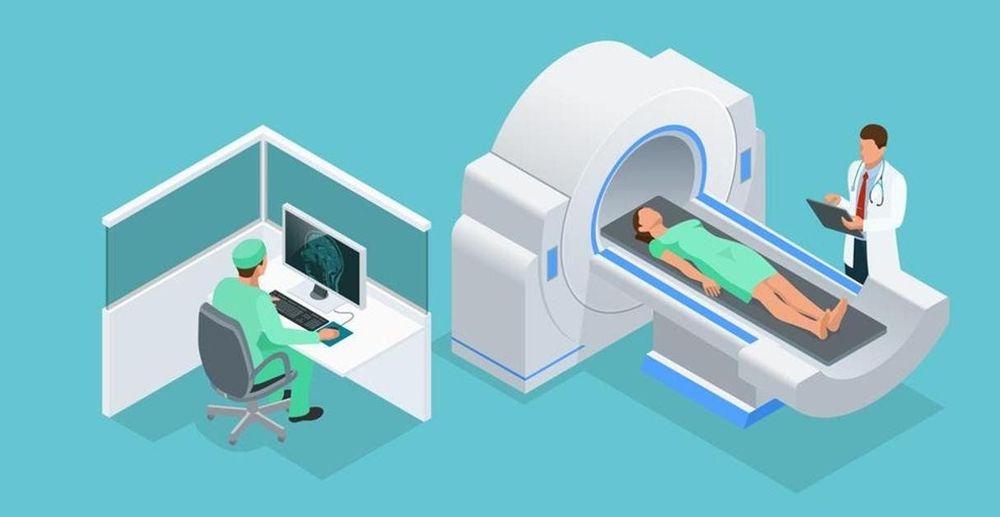
In order to identify patients at high risk of ischemic stroke and cerebral edema, it is necessary to use clinical data, including embolism. Imaging tests are used to increase the efficiency of diagnosing the risk of cerebral edema after stroke. Specifically:
Brain CT scan without contrast: The first choice is useful to diagnose and monitor patients with cerebral edema due to cerebral hemisphere or cerebellar infarction. Follow-up CT scans during the first 2 days are useful to identify patients at high risk of developing symptomatic edema. The first 6 h CT scan showing hypoattenuation, focal area of NM≥ 1/3 MCA, and midline deviation are useful markers in predicting cerebral edema. 6 h MRI DWI volume measurement is helpful; if V>80ml predict the progression will be very fast

Chụp CT scan não không cản quang là một trong những phương pháp chẩn đoán phù não
3. Treatment of cerebral edema after stroke
Patients can be given osmotic therapy for stroke cases that have worsened, at risk of cerebrovascular edema. Patients should also undergo decompression craniotomy for patients <60 years of age with an MCA infarction that worsens within 48 h despite maximal medical therapy. The effect of subsequent decompression is unknown but warrants strong consideration. Simultaneously, decompression suboccipital craniotomy should be performed in patients with cerebellar infarction whose neurological deterioration has worsened despite maximal medical therapy. The doctor's appointment should be based on the criteria for the indication that craniotomy to relieve pressure through impaired consciousness and brain edema is reasonable to bring maximum treatment effect to the patient.
Vinmec International General Hospital with a system of modern facilities, medical equipment and a team of experts and doctors with many years of experience in neurological examination and treatment, patients can completely peace of mind to examine and treat neurological diseases in general and cerebral edema after stroke in particular at Vinmec.
To register for examination and treatment at Vinmec International General Hospital, you can contact Vinmec Health System nationwide, or register online HERE.


