This is an automatically translated article.
The article is professionally consulted by Master, Doctor Nguyen Van Phan - Head of Interventional Imaging Unit - Department of Diagnostic Imaging and Nuclear Medicine - Vinmec Times City International General Hospital.Digital hepatic angiography with background erasure is an interventional procedure using iodinated contrast agent to image the visceral artery, common hepatic artery, separate hepatic artery, right hepatic artery, left hepatic artery and fecal branches lobes, subsegments in the liver. Digital liver angiography erases background to help diagnose liver tumors with angiogenesis, aneurysms and vascular malformations, thereby providing appropriate treatment.
1. Indications, contraindications when digitized hepatic artery angiography?
1.1 Designation
● Patients with primary liver cancer are indicated for hepatic artery embolization with chemotherapy in combination with Lipiodol (TACE) or embolization with chemo-carrying microspheres, radioactive microspheres, or embolization to reduce the size of the vessel. Preoperative tumor size, radiofrequency ablation.● Primary liver cancer, or secondary angiogenesis but no indication for surgery.
● Large liver cancer tumors burst causing abdominal bleeding, affecting the patient's life and needing to be embolized.
● Large benign liver tumors cause pain symptoms or are at risk of rupture .
● Plug the hepatic artery aneurysm with a metal helix, malformation of the hepatic artery - portal vein.
● Acute abdominal bleeding after traumatic liver rupture.

Bệnh nhân bị ung thư gan nguyên phát có chỉ định chụp để điều trị các khối u gan trước phẫu thuật
1.2 Contraindications
● Severe and uncontrolled clotting disorders (prothrombin <60%, INR > 1.5, platelet count < 50 G/l): Need to be adjusted before intervention● Severe cirrhosis: Child pugh C
● Blood portal vein block
Grade IV severe renal failure
Allergy to iodinated contrast media
Pregnant women
2. Process of digitizing liver angiography with background erasure
2.1 Personnel
2 interventional radiologists 1 nurse 2 radiology technicians 1 anesthesiologist if the patient is uncooperative or the child is under 5 years old. 1 anesthesiologist2.2 Vehicles and machines
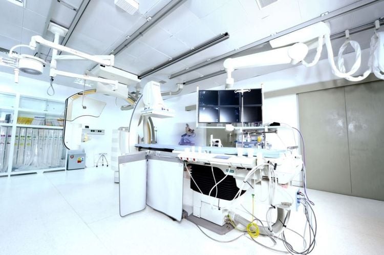
● Digital erasure angiography (DSA)● Specialized high-pressure contrast agent pump.
Pacs image storage system.
● Lead jacket, apron, lead collar, x-ray shielding glasses.

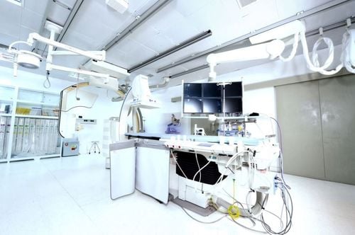
Máy chụp mạch số hóa xóa nền (DSA)
2.3 Implementation steps
● Patients and guardians are explained about the procedure, benefits, risks of complications - possible complications. Sign a commitment to do the procedure, commit to using contrast agents containing iodine.● Fasting, drinking before 6 hours if anesthesia; 4 hours before anesthesia. You can drink water but not more than 50ml of water.
● At the intervention room: the patient lies on his back, installing a monitor to monitor breathing, pulse, blood pressure, electrocardiogram, SpO2.
● Disinfect the femoral artery puncture area, cover with a vascular intervention towel set.
● Local anesthesia of femoral artery puncture with Lidocaine 2%
● Fetal artery puncture. In special cases, a radial puncture can be performed if femoral artery puncture is not possible. Place the artery opening kit.
● Insert the catheter into the visceral angiogram to evaluate the overall hepatic artery, splenic artery, and gastroduodenal artery.
Superior mesenteric angiogram to evaluate the portal vein system.
● Identify the damaged blood vessel, use the microcatheter to thread the superselectively into the vascular peduncle supplying blood to the tumor.
● Superselective embolization of the tumor with a chemical suspension combined with Lipiodol (TACE) or embolization with chemical-carrying microspheres, radioactive microspheres until the entire tumor is completely occluded.
● Take a scan to check the occlusion of the feeding pedicles, continue selective embolization if there are any. Finally, the vascular node feeds the tumor with bio-foam (gelfoam).
● Remove the catheter and open the catheter into the lumen.
● At the end of the procedure, compress the femoral artery or close the femoral artery.
● The patient lies on the hospital bed, immobilizes the leg on the side of the puncture, the nurse will monitor for bleeding complications ~ 8 hours if the bandage is pressed, ~ 2 hours if the femoral pulse is closed.
● Monitor pulse, temperature, blood pressure, respiratory indicators.
● Use pain relievers and antiemetics in case of need.
● Maintain drugs that support liver function.
Dr. Nguyen Van Phan is an interventional radiologist and radiologist, with extensive experience in diagnosing and performing vascular interventions such as brain aneurysm implantation, brain stenting, and vascular malformations. cerebrovascular brain; embolization to treat hemoptysis; treatment of liver tumors, uterine fibroids, benign prostatic hypertrophy, stem cell transplantation for treatment of cirrhosis, congenital biliary atrophy, Type II diabetes... Non-vascular interventions such as high-frequency ablation frequency (RFA) treatment of liver tumors, benign thyroid tumors; aspiration and drainage of abscesses, drainage and stenting of biliary tract, urinary kidney; aspiration and biopsy of the breast, thyroid, lymph nodes, software, biopsy of liver tumors, lung tumors, bone tumors ...
All questions need to be answered by specialists as well as customers. If you want to be examined and treated at Vinmec International General Hospital, you can contact Vinmec Health System nationwide or register online HERE.
SEE MORE
The procedure of computed tomography in the hepatic artery chemotherapy node Digital erasure of the background and liver biopsy through the jugular vein in Liver CT: What you need to know: What you need to know


