This is an automatically translated article.
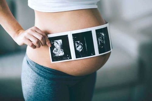
Placenta is a very important part, providing oxygen and nutrients for the developing fetus. Ultrasound is a safe diagnostic method that can detect placental diseases early, so that early intervention can be taken to prevent complications and help the fetus develop healthy.
1. Placenta cake
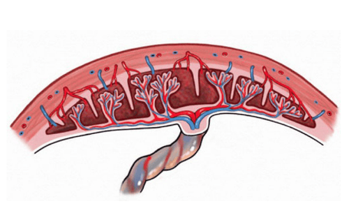
Placenta is the place where many blood vessels come and usually attaches mainly to the fundus of the mother's uterus. The placenta is round about 15cm in diameter, weighs 1⁄6 the weight of the fetus and is 2.5-3cm thick. Each placenta consists of 15-20 segments, between the segments are small grooves. The placenta is divided into 3 parts including:
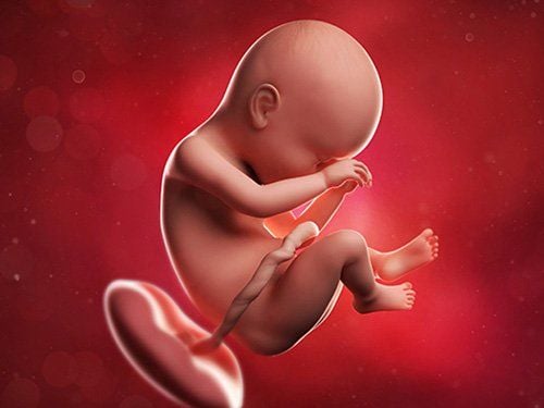
Basal layer: The part in contact with the uterine muscle; Buffer layer: Is the part of the placenta towards the fetus, this is the adhesion of membranes to the placenta; Organize each other. The placenta has the function of providing oxygen and nutrients to help the fetus develop, transport substances from the mother to the fetus and then waste products from the fetus to the mother. The placenta has the ability to make nutrients reach the fetus faster and more efficiently. In addition, the placenta also protects the fetus from the risk of many factors from inside the mother's body and from the environment.
2. Normal placental ultrasound
At the 8th week of pregnancy, layers of trophoblastic cells surround the amniotic sac creating a thick echogenic border around the gestational sac. Weeks 10-12 have differentiated placenta. In the first trimester, the amniotic membrane separates from the chorion. Then the amniotic sac enlarges and the amniotic membrane merges into the chorionic cavity to form an ectopic oocyte.
By the 4th month, the uterus grows and the placenta is localized, the thickness of the placenta increases, from which it is possible to predict the position of the placenta in the future. The thickness of the placenta corresponds to the gestational age as follows:
20 weeks pregnant : 20mm placental thickness; 30 weeks pregnant: The thickness of the placenta is 30mm; 40 weeks pregnant: The thickness of the placenta is 40mm. However, in the case of polyhydramnios, the placenta is really thick, but the amniotic fluid is compressed, and it cannot develop.
The maturity of the placenta is classified as follows:
Grade 0: The plate is smooth and flat, the placental tissue is homogenous, there are no calcifications in pregnancy I - II; Grade 1: Wavy, undulating pad, echogenic placental tissue scattered with calcified highlights; Grade 2: Basal plate has calcified, comma-enhanced echoes from the placental plate into the placental tissue. This corresponds to the calcification of the septum; Grade 3: Buffer imprint, clearly distinguishing the plate and the bottom plate, the placenta has calcified circles, calcification of the bottom plate appears. The gain line starts from the accompaniment to the bottom. Classification of position of placental attachment:
Placental attachment anteriorly: Group I: The upper edge of the placenta attaches to the posterior 1⁄4; Group II: The upper edge of the placenta is attached to the anterior-superior 1/4; Group III: The upper edge of the placenta is attached to the anterior and inferior 1/4. The placenta attaches posteriorly: Group I: The upper edge of the placenta attaches to the anterior and superior 1⁄4; Group II: The upper edge of the placenta is attached to the posterior 1⁄4; Group III: The upper edge of the placenta attaches to the posterior 1⁄4 below.
Bánh nhau là nơi chứa nhiều mạch máu đến và thường bám chủ yếu trên đáy tử cung của người mẹ
3. Ultrasound of placental diseases
3.1 Placenta praevia Placenta previa occurs when the placenta attaches to or near the opening of the cervix or completely covers the opening of the uterus. The case that completely covers the hole in the uterus by the placenta is called the placenta central previa. In cases where the placenta does not completely cover the hole in the uterus, it is called a semi-central placenta previa.
Patient will have symptoms of vaginal bleeding occurring during pregnancy and 3rd trimester including:
No abdominal pain; Bright red blood; Fetal heart can be heard clearly; Abdomen without contractions; There were no abnormalities of coagulopathy. Ultrasound images in the pathology of placenta previa include:
Placenta previa is divided into 4 types depending on whether the placenta attaches to the front or back of the uterus; Placenta striker front and back. 3.2 Placental abruption Placental abruption is common due to trauma and has a history of placenta previa, short umbilical cord, high blood pressure, vascular disease,... Pregnant women often present with symptoms of pain and bleeding. vaginally or not, uterine wall distension and signs of preterm labor and shock following a direct or indirect trauma.
On ultrasound images appear post-placental hematomas, which can be central or at the edge of the placenta. In addition, the placenta is thickened but only 50% can be seen on ultrasound.
3.3 Placenta Comb Placenta is an abnormally adhered placenta where hairs can penetrate the lining of the uterus into the muscularis layer of the uterus. Placenta comb usually accompanies the placenta previa in 5-10% of the cases of placenta previa. The common cause of placenta previa is a scar in the uterus such as a cesarean section, fibroid removal, multiple abortions or after a manual placental removal procedure after giving birth,... The next time you get pregnant, the placenta can stick to old scars and can cause severe bleeding during pregnancy.
Placenta accreta has 3 levels depending on the adhesion of the placenta through the uterine muscle, including:
Placenta accreta: Placenta adheres to the uterine lining without invading the uterine muscle; Placenta accreta: The placenta attaches to the uterine muscle but has not yet invaded the serosa; Placenta percreta: The placenta attaches through the uterine myometrium and through the serosa. The method of diagnosis of placenta accreta based on doppler ultrasound will clearly see the blood vessels located in the uterine muscle. In addition, 2-dimensional ultrasound is possible, but difficult to diagnose.
In summary, ultrasound is a safe and commonly used diagnostic method in obstetrics. Application of ultrasound in the diagnosis of placental disease can detect abnormalities in the position of attachment, size... of the placenta. From there, early and appropriate interventions can be given. Therefore, pregnant women should pay attention to go for regular antenatal check-up on time, and when there are abnormalities such as vaginal bleeding, fatigue, etc., they need to go to a medical facility immediately for examination and timely treatment. .
Chẩn đoán nhau cài răng lược dựa vào siêu âm doppler sẽ thấy rõ các mạch máu nằm trong cơ tử cung
Ultrasound is a quick and effective way to detect and diagnose some placental diseases. If you suspect that you have placental disease, you can come to Vinmec International General Hospital System. As one of the leading prestigious hospitals in the country, Vinmec uses the most modern generations of color ultrasound machines today in examining and treating patients. One of them is GE Healthcarecar's Logig E9 ultrasound machine with full options, HD resolution probes for clear images, accurate assessment of lesions. In addition, a team of experienced doctors and nurses will greatly assist in diagnosing and early detection of abnormal signs of the body in order to provide timely treatment for placental disease.
Please dial HOTLINE for more information or register for an appointment HERE. Download MyVinmec app to make appointments faster and to manage your bookings easily.