This is an automatically translated article.
Head and neck anatomy image is a scientific study. Thanks to anatomical images, it is possible to determine the correct position of organs. This is also the basis for scientific research.
1. How to determine the head and neck area?
In medicine, each body part is clearly divided according to characteristics and functions. Anatomy of the head and neck area is to separate the surrounding area to help medical research. The head and neck region is the area of the organs from the head to the neck. Based on that, doctors can define the treatment area for the patient quickly.
2. Parts of the head and neck area
2.1. The skull The skull is the part of the brain that protects the brain when the head is hit. But when the impact is strong, it causes damage and is evaluated as a traumatic brain injury. Carrying out the anatomy of the human skull, it is possible to find a few other sets of paired parts such as:
Frontal bone Nasal bone Lacrimal gland bone Upper jaw bone Nasal cortex Lower jaw Occipital bone Parietal bone Temporal bone Temporal bone Cane bones Cheekbones. These parts are linked together into a unified whole with an elastic structure through the joints for separation. From an analytical point of view, between the pieces of bone grafted together, there is a joint like a fixed suture.
2.2. Nose in the head and neck region The nose is the protruding part of our face listed in the sense of smell. The nose can be used to smell and exchange air. There are bones and cartilage in the nose that help shape the septum to allow air to flow out. Behind the nose is the nasal cavity. Because the nose has 2 sides, there are 2 nasal cavities and nasal septums.
At each nasal cavity there are 3 structures divided into 3 compartments, so they are collectively called the nasal cavity. The sinuses are distributed along the bridge of your nose and in your head and skull. At the nose there are also arteries that pass through them connecting the nose with the brain and facial organs.
2.3. Eye Parts On the skull there are 2 deep hollows where the eyes develop. The eyeball will hover in that bone cavity in an orbit so that it is convenient for people to make eye contact and see many different angles. The anatomy of the eyeball is quite complicated because it has the ability to help your eyes widen. In addition to the lens that helps the eye to see objects, the composition of the eyeball also has a number of other substances that need further analysis for identification.
The pupil is the black part controlled by the iris. The pupil of the eye receives the transmitted light, and the reflected point is the retina. This is a reflection mechanism in physics that makes it possible for the eye to see everything in light conditions. In addition, there are eyelids, conjunctiva, conjunctiva, and tear glands that contribute to the protection of the eyes.
The ophthalmic artery connects to the optic nerve, oculomotor... They are also linked to the skull to facilitate control of eye behavior. At the same time, it is a wire that transmits information that helps the brain perceive what the eye is seeing.
2.4. Ear in the head and neck region The ear has 3 parts: the outer ear, the middle ear and the inner ear. Each part has different functions from capturing sound, transmitting sound to receiving sound. The arteries in the ear are connected to the nose and throat. They are all directed to the brain to be able to provide the fastest sensory information and give reflex commands to the muscles when there is danger.
2.5. The mouth is the part of the head and neck region The oral cavity includes teeth, tongue, cheeks, tonsils, pharynx... This is the first part to digest food. They have the function of receiving and crushing food before it is taken to the intestines. The mouth has two vestibule parts located between the teeth and lips, and the oral cavity is behind. The arteries here contribute to the control of the jaw muscles to work smoothly. They combine with facial muscles to help people express emotions and perform the act of chewing food.
2.6. Teeth in the head and neck region Teeth belong to the oral cavity but are classified as dental. They have a complex anatomical structure and are divided into two stages in human life. Children under 3 years old have teeth called baby teeth, after the loss of baby teeth, new replacement teeth are counted as adult teeth. These teeth are classified by function as incisors, molars, and canines.
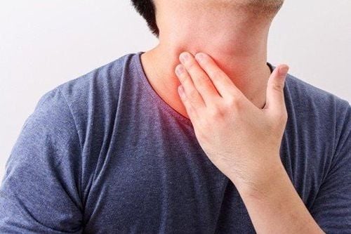
2.7. The most complicated part of the neck is probably the neck area. Here there are many tight muscle bundles and tight nerve fibers. Behind the inside of the neck are the thyroid, larynx, and pharynx. There are 4 arteries that pass through the neck you need to know that are common carotid artery, internal carotid artery, sacral carotid artery, external carotid artery.
3. Medical images can clearly show the anatomy of the head and neck area
Anatomical images of head, face and neck muscles have great medical significance. Anatomical imaging is a research facility that helps doctors to clearly identify each part. This image can help make the diagnosis easier when surgery is needed, especially with current laparoscopic techniques.
In teaching, the anatomical model of the head, face and neck is the basis for medical students to understand. It is also a simulator to help students learn and practice surgery.
Head and neck muscle surgery can also help improve facial tone. In plastic surgery, the adjustment of facial morphology needs to be based on or based on pre-existing facial muscle blocks to perform.
The image of muscle anatomy of the head, face and neck has great practical significance. In addition to spreading knowledge to medical students, this is also a model to support doctors in research to be able to treat many dangerous diseases in the head and neck region.
Please dial HOTLINE for more information or register for an appointment HERE. Download MyVinmec app to make appointments faster and to manage your bookings easily.
Reference sources: oncolink.org, kenhub.com


