
This is an automatically translated article.
The article was professionally consulted by Doctor Tran Thi Diem Trang - Respiratory Internal Medicine Doctor - Department of Medical Examination & Internal Medicine - Vinmec Central Park International General Hospital. The doctor has more than 10 years of experience in treating respiratory diseases.Acute respiratory distress syndrome in adults is a manifestation of severe disturbance of blood oxygen exchange, severe cases are common in patients with no history of cardiovascular and respiratory disease.
1. What is acute respiratory failure?
Acute respiratory failure is a condition in which the lungs suddenly fail to function properly for gas exchange, causing hypoxemia, with or without hypercapnia.Hypoxemia alone is not necessarily milder than hypoxia with hypercapnia, sometimes more severe as in acute respiratory distress syndrome in adults.
(ARDS: adult respiratory distress syndrome).
The elderly are susceptible to respiratory diseases, including respiratory failure in the elderly.
Acute respiratory failure is the most common emergency, requiring immediate intervention. In fact, acute respiratory failure can be divided into 2 types:
Severe: Major drug intervention, which can be resolved with medication or some minor procedures. Critical type: must intervene immediately by procedures then use drugs or must be used in parallel (intubation, balloon compression, mechanical ventilation...)
2. Causes of acute respiratory failure
Pulmonary causes: bronchopulmonary infection, pulmonary embolism, pneumothorax, bronchial asthma, acute bronchial obstruction... Extrapulmonary causes: laryngotracheal obstruction, pleural effusion lung injury, thoracic injury, thoracic injury, respiratory muscle injury, central nervous system damage.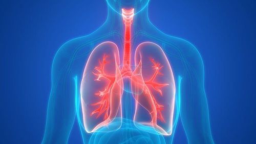
Suy hô hấp cấp tính có thể do nguyên nhân tại phổi
3. Symptoms of acute respiratory failure
3.1 Clinical symptoms Shortness of breath : Lack of blood oxygen with increased or not increased PaCO2 also causes shortness of breath.Breathing rate: May increase, often with accessory respiratory muscle contractions as in bronchopneumonia. May be reduced, without traction due to central respiratory paralysis in barbituric poisoning. Ventilator must be indicated immediately because the breathing rate will slow down.
Respiratory amplitude :
Decreased in bronchopneumonia, cobra bite, polio, Guillai-Barre syndrome. Increased in acute respiratory distress syndrome, pulmonary embolism. Blue-violet:
On lips and fingertips. When reduced Hb is above 5g/100ml, SaO2 is below 85g/l. The extremities are still hot, unlike shock. No cyanosis if anemia. There is no cyanosis but purplish red, sweating if the PaCO2 is increased as much as in the acute exacerbation of chronic bronchitis. Often accompanied by a drumstick finger. Cardiovascular disorders:
Rhythm: usually sinus tachycardia or tachycardia (flutter, atrial fibrillation or junctional tachycardia). Erratic vibration is the final manifestation. Blood pressure rises or falls: In the first stage, blood pressure is often elevated, then gradually lowers, requiring immediate intervention (ballooning, endotracheal intubation, sputum suction, mechanical ventilation). Cardiac arrest due to severe hypoxia or excessive PaCO2 requires immediate medical attention. Can recover quickly if intervention before 5 minutes. Nervous disorders and consciousness: The brain consumes 1/5 of the total oxygen in the body. So the brain suffers the earliest consequences of hypoxia and hypercapnia.
Nervous disorders: Struggling, confusion, loss of tendon reflexes.
Disorders of consciousness: Lethargy, lethargy, coma.
3.2 Paraclinical symptoms Pulmonary angiography:
On-site chest x-ray is required for all patients with acute respiratory failure. However, it is difficult to perform an electrocardiogram in a rapidly breathing, bed-ridden patient or a patient in a coma due to barbituric poisoning who is on a ventilator.
Prepare for a chest x-ray in the following manner: place the patient on a ventilator with 100% hyperventilation and oxygen or squeeze the balloon through a mask for 20 minutes. Oxygen saturation will increase rapidly, breathing will slow down or stop completely within a few minutes.
Blood gas tests:
Blood gas tests include:
SaO2 (arterial blood oxygen saturation): Normal is 95-97%. SaO2 below 85% is purple. PaO2 (arterial blood oxygen pressure): Normal in young people is 95-96 mmHg, in people over 60 years old is 78 mmHg. In acute respiratory failure, PaSO2 falls below 40 mmHg (8kPa). PaCO2 (arterial blood CO2 pressure): Normally equal to 40 mmHg, can reach 80 mmHg (10.7 kPa) in acute respiratory failure or more. PaCO2 is increased in alveolar hypoventilation. Testing for blood gases allows to classify acute respiratory failure into two main groups.
Group I: Hypoxemia without elevation of CO2
PaPO2 falls below 8 kPa. Normal or low PaCO2 accompanied by: Respiratory alkalosis due to alveolar hyperventilation, or metabolic acidosis due to increased lactic acidosis. Example: ARDS, shock. Group II: Decreased alveolar ventilation
PaO2 decreased. PaCO2 increased. Often accompanied by respiratory acidosis or mixed acidosis (associated with hyperlactic acidemia). Example: Respiratory paralysis, obstructive bronchitis. Group I only have hypoxemia does not mean a milder prognosis than group 2. In acute respiratory distress syndrome with acute respiratory failure, oxygen only diffuses through the alveolar wall in the healthy area, so oxygen with FiO2 = 1 sometimes it does not bring PaO2 back to normal.
4. Diagnosis of acute respiratory failure
The definitive diagnosis of acute respiratory failure is usually easy, it is a clinical diagnosis. Determining the type of acute respiratory failure can be more difficult because it relies on testing. The best solution is to determine the cause and then find out the pathogenic mechanism to decide the treatment attitude.Differential diagnosis is also important to avoid incorrect management:
Hyperventilation (not dyspnoea) is present in metabolic acidosis, aspirin poisoning, brain stem damage. Cheyne-Stokes breathing is common in cases other than respiratory failure such as: heart failure, kidney failure, cerebrovascular accident. Sometimes seen in opium poisoning (hereinafter indicated mechanical ventilation). Cyanosis and shortness of breath may be caused by pericardial effusion causing cardiac compression, vitamin B1 deficiency (usually loss of tendon reflexes). Encephalopathy due to acute respiratory failure can be confused with encephalitis with acute respiratory failure. Malignant malaria often has pulmonary complications that make the disease worse. Respiratory resuscitation measures need to be taken immediately, the more urgent the patient's survival.

Chẩn đoán quyết định suy hô hấp cấp thường là dễ, đó là một chẩn đoán lâm sàng
5. Treatment of acute respiratory failure
Treatment of patients with ARDS requires attention to (1) identification and treatment of the underlying disease; (2) Mechanical ventilation with lung protection mode; (3) Fluid balance (4) minimizing complications of treatment and procedures; (5) prevent venous thrombosis, gastrointestinal bleeding, aspiration, avoid excessive sedation, avoid infection (6) provide adequate nutrition.Identify and treat causative diseases Identifying and treating causative diseases is very important in the management of ARDS. Only by eliminating the cause of ARDS can the patient emerge from respiratory failure.
Mechanical ventilation: Patients with severe ARDS with increased work of breathing and progressive hypoxemia require mechanical ventilation. The requirement of mechanical ventilation is to ensure adequate arterial oxygenation and minimize the risk of ventilator-associated complications. Fluid balance: in essence, ARDS is acute pulmonary edema. If the fluid balance is positive, along with increased vascular permeability in ARDS and hypoproteinemia are favorable factors for the development of acute hemodynamic pulmonary edema. Maintain a normal or low left atrial filling pressure to minimize pulmonary edema and prevent further reduction of arterial oxygenation and lung elasticity, improve lung mechanics, shorten breathing time machine and hospital stay, and associated with lower mortality in ICU and surgical patients. Therefore, reducing left atrial filling pressure with fluid restriction and diuretics is important in the treatment of ARDS. Caution should be exercised in patients with hemodynamic instability and hypoperfusion of vital organs such as the kidney. Corticosteroids Many studies have investigated corticosteroids in the treatment of both early and late ARDS to reduce lung inflammation. Current evidence does not support the use of high-dose corticosteroids to treat ARDS,. However, there are specific situations where corticosteroids can be used with caution when the underlying cause responds to corticosteroid treatment (eg. such as cryptogenic organizing pneumonia).
Other Treatments In clinical trials, most pharmacological treatments have shown disappointing results.
- Inhalation of nitric oxide (NO) may transiently improve oxygenation but neither mortality nor duration of mechanical ventilation.
- Beta 2 agonist (salbutamol): theoretically reduces alveolar capillary membrane permeability, initiates recovery earlier, evidence suggests that beta 2 agonist does not reduce time to mechanical ventilation and rate In-hospital mortality
- Surfactant: may improve lung function, does not improve mortality and duration of mechanical ventilation
- Ketoconazole, thromboxane A2 and leukotriene synthesis inhibitor, not proven to prevent prevent ARDS, do not improve mortality and duration of mechanical ventilation.
- Lisofylline: no difference in mortality and duration of mechanical ventilation
- Sivelestat: inhibits neutrophil elaspase, does not improve long-term mortality.
Please dial HOTLINE for more information or register for an appointment HERE. Download MyVinmec app to make appointments faster and to manage your bookings easily.