This is an automatically translated article.
Phyllodema is a type of tumor that accounts for about 0.3% - 0.5% of all malignancies of the mammary gland. It is common in non-menopausal women between the ages of 40 and 50 and in men, although it is very rare.
1. What is U phyllode?
A phylloderm tumor (or chlorophyll tumor) is a growth of normal cells that develops into a hard lump in the breast. These phyllodes tumors have a leaf-like structure, well-defined boundaries, and are composed of connective tissue and epithelium. Usually when making the diagnosis, phyllodes tumors will be quite large (between 4 and 5 cm).
Cystic tumors can be:
Benign (non-cancerous) tumors; Malignant tumor (cancerous); Borderline tumor (located at the border between benign and malignant). According to statistics given by experts, the majority of breast chlorophyll tumors are benign tumors with slow progression, so the prognosis after treatment is very positive. Tumors of phyllodes (phyllodes tumor) are usually treated with surgery. Most patients with borderline phyllodes tumor will have no further problems after treatment.
SEE ALSO: How is mammary syphilis formed and is it dangerous?
2. Signs of phyllodes tumor
The most common signs of breast phyllodes tumor are easy to feel, patients can feel them when pressing on the skin of the breast. They will usually form in the upper quadrant of the breast. During the first few weeks or months, the size of the phylloderm can reach as little as 2 to 3 centimeters. Although the average size of this breast phyllodes tumor is about 4 cm, they can be many times larger. Approximately 20% of phyllodes tumors exceed 10 cm in diameter. This type of tumor does not cause pain to the patient.
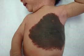
Because the phyllodes tumor can extend into the veins under the skin, that area of the breast will appear blue. Whether a patient's phyllodes tumor is benign or malignant, it is possible for ulcers to form on the skin of the breast (however rare). Phyllodes tumors are likely to form in both breasts, but this is very rare.

Dấu hiệu phổ biến nhất của khối u diệp thể vú rất dễ cảm nhận
3. Subjects usually diagnosed with phyllodes tumor
Basically, breast syphilis can appear at any age but will be most common in women between the ages of 40 and 50 who have not gone through menopause. In addition, although very rare, phyllodes tumors can occur in men.
Benign chlorophyll tumors are more common than borderline and malignant tumors. Usually, phyllodes tumor will be treated by surgical option.
SEE ALSO: Identify some common benign diseases in the mammary glands
4. Diagnosis of phyllodes tumor
Cases of phyllodes tumor are very rare, they only account for about 0.3% to 0.5% of breast tumors in women. Therefore, this causes many difficulties for doctors in diagnosing phyllodes tumors. In addition, because phyllodes tumors may resemble fibroadenomas, a differential diagnosis between the two conditions is required. There are two main differences between tumor types:
Growth rate: Breast phyllodes tumor will tend to grow faster than fibroadenoma. Age: Usually phyllodes tumors tend to start when people are in their 40s. Fibroids are more common in their 20s and 30s. These differences can help your doctor make a primary diagnosis. corpse. As with other tumors, the diagnosis of a phylloderm tumor may include the following tests:
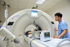
Mammogram : The imaging shows a round mass with well-defined edges. Round lobes may appear in the tumor in some isolated cases. Magnetic resonance imaging (MRI) or ultrasound: These test images will provide doctors with more information, supporting the diagnosis. Biopsy takes a sample of the tumor and examines it under a microscope: This is the only way to know if this growth is a phyllodema. There are two types of biopsies: core needle biopsies and excisional biopsies. Besides, not only can help confirm tumor type, biopsy can also help determine whether this phylloderm is benign, malignant or borderline.

Chụp cộng hưởng từ vú chẩn đoán khối u phyllode
5. How to treat phyllodes tumor?
5.1. Surgery The only way to treat phyllodes is to completely remove the tumor. This treatment is aimed at preventing the formation of new tumors as well as stopping complications caused by existing tumors (whether benign, malignant or borderline). The surgeon will remove the tumor and at least 1 cm of surrounding tissue to reduce the chance of the tumor growing back. In cases where the tumor is cancerous, the area of surrounding tissue that needs to be removed may be more.
If a cancerous syphilis tumor recurs, your doctor may recommend a mastectomy (removal of part or all of the breast). In addition, your doctor may recommend a combination of radiation therapy, chemotherapy, or both. Although benign phyllodes tumors will not spread beyond the breast, treatment is still needed to prevent them from growing larger.
After removal of a breast phylloderm, pain may be felt at the surgical site, but subsequent complications are rare. Benign breast chlorophyll tumors are less likely to recur than malignant tumors. Patients will be monitored for recurrence, usually between 1 and 2 years after removal.
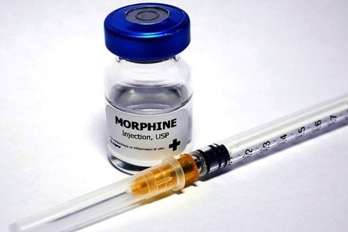
5.2. Radiotherapy For patients with benign phyllodes tumors, there is no indication for radiation therapy. The doctor will prescribe adjuvant radiation therapy for cases of phylloderm tumors in borderline or malignant form.

Chỉ định xạ trị bổ trợ cho các trường hợp khối u phyllode ở thể giáp biên hoặc thể ác tính
5.3. Chemotherapy Because of the small number of patients with breast phylloblastoma, the data are limited, whereby the role of systemic chemotherapy remains unclear. Indications for adjuvant chemotherapy for malignant tumors are still controversial.
Although, phyllodes tumor has a very good prognosis because it is usually benign, but if not diagnosed and treated promptly, even benign tumors can cause complications for patients. Therefore, if anyone notices that he or she has abnormal breast-related changes, they should visit hospitals with breast screening specialties such as Breast Screening Center at Vinmec Times City for examination as soon as possible.
Breast cancer is a common cancer in women, it is estimated that every year around the world, many women die from this disease. Accordingly, if breast cancer is detected early, the prognosis for treatment is very high, even if it is completely cured and not recurred. Therefore, breast cancer screening is essential, especially for subjects with high risk of breast cancer such as breast tumors,...
Currently, General Hospital Vinmec International has been implementing a breast cancer screening package for women of different ages. At Vinmec, there is a full range of necessary medical equipment, a system of modern medical machines to perform methods of examination, diagnosis, testing, imaging, and treatment of breast cancer at many stages. . Accordingly, the breast cancer examination and treatment process at Vinmec is performed by a team of highly qualified doctors at the Breast Screening Center in Vinmec, who have undergone training and been granted technical certificates. can be handled quickly and efficiently. Therefore, when using Vinmec's breast cancer screening and early detection package, customers can detect cancer even when there are no symptoms.
Please dial HOTLINE for more information or register for an appointment HERE. Download MyVinmec app to make appointments faster and to manage your bookings easily.