This is an automatically translated article.
The article is professionally consulted by Master, Doctor Nguyen Viet Thu - Doctor of Radiology and Nuclear Medicine - Department of Diagnostic Imaging and Nuclear Medicine - Vinmec Times City International HospitalUltrasound is an imaging technique that uses ultrasound waves (high frequency sound waves) to build and reconstruct images of the internal structures of the body. Ultrasound is one of the most widely used diagnostic and therapeutic methods in medicine.
1. Learn the concept of ultrasound
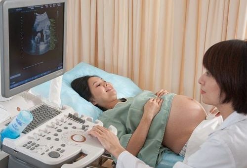
Ultrasound is a subclinical method that effectively supports doctors in diagnosing and monitoring patients. This is a popular, effective and safe method, but requires a prescription and recommendation from a doctor.

Siêu âm là một phương pháp cận lâm sàng hỗ trợ đắc lực cho các bác sĩ trong việc chẩn đoán và theo dõi bệnh nhân
Ultrasound is used to survey many important parts and organs in the body such as: abdomen, obstetrics, cardiology, gynecology, mammary gland, thyroid gland.... and technical support for other medicine.
2. What does the patient need to prepare for the ultrasound?
Preparation for ultrasound depends on the patient's location to be examined. There are some types of ultrasound, which the patient does not need to prepare in advance. But there are some types, patients need to abstain from certain foods, drinks or hold urine for a few hours before the ultrasound. For example, for many gallbladder ultrasounds, the patient needs to fast before going for the ultrasound.
Doctors recommend that patients should wear comfortable, loose clothes because they may have to remove their clothes during the ultrasound.
3. Procedure performed in ultrasound
Step 1: After the patient is ready for the ultrasound, the doctor will apply a gel to the area to be examined. The effect of this gel helps the transducer make sure contact with the body, limiting the air between the transducer and the patient's skin.
Step 2: The doctor uses a transducer that both transmits and receives ultrasound waves close to the patient's skin and scans it on the areas of the body to be examined. The procedure is very gentle and the patient will not feel any pain or discomfort.

Quy trình siêu âm gồm 3 bước
4. Techniques and some terminology used in ultrasound
2D ultrasound - 2D ultrasound is the most used method in current ultrasound procedures.
3D ultrasound - 3D ultrasound is often used in fetal ultrasound, thyroid ultrasound
Continuous wave Doppler ultrasound to ultrasound blood vessels
Types of echocardiography, general abdominal ultrasound, ultrasonography transvaginal ultrasound, mammogram...
5. Advantages of ultrasonic technique

Kỹ thuật siêu âm có nhiều ưu điểm
Helps to examine most of the diseases such as: tumors, inflammation, deformities... at positions such as abdominal cavity, pelvis, liver, bile, kidney... Assess the development of the fetus, Especially with 3D and 4D ultrasound, doctors can evaluate most of the morphological abnormalities of the fetus. Accurately assess the degree of effusion of the pleura, pericardium... Ultrasound also accurately assesses the size and location of stones in the diagnosis of kidney stones, bladder stones, urethra. Depending on the quality of the ultrasound machine and the diagnostic ability of the doctor, the patient can be diagnosed quickly. Currently, there are many medical facilities that apply ultrasound technology in the early diagnosis and treatment of diseases. Vinmec International General Hospital system has been using the most modern generations of color ultrasound machines today in examining and treating patients. One of them is GE Healthcarecar's Logig E9 ultrasound machine with full options, HD resolution probes for clear images, accurate assessment of lesions. In addition, a team of experienced doctors and nurses will greatly assist in the diagnosis and early detection of abnormal signs of the body in order to provide timely treatment.

Hệ thống Bệnh viện Đa khoa Quốc tế Vinmec đã và đang sử dụng các thế hệ máy siêu âm màu hiện đại nhất hiện nay


