This is an automatically translated article.
The article is professionally consulted by Master, Doctor. Doan Xuan Sinh - Head of Diagnostic Imaging Department - Department of Diagnostic Imaging - Vinmec Hai Phong International General HospitalContrast-enhanced ultrasonography (CEUS) is indicated in cases where it is necessary to evaluate the kinetics of tumors, especially liver tumors. Support in percutaneous intervention under the guidance of ultrasound and evaluate the effectiveness after treatment (after radiofrequency ablation, alcohol injection, hepatic embolization...)
1 What is Contrast-Enhanced Ultrasound (CEUS)?
Contrast-enhanced ultrasound (CEUS) is the application of an ultrasonic contrast agent to conventional ultrasound, based on the many ways in which sound waves are reflected from the interfaces between substances, which can be: the surface of a microbubble or a more complex structure. Usually gas-filled microbubbles are injected intravenously into the circulatory system. Microbubbles have high echogenicity, so they are able to reflect sound waves. Due to the great difference between the echogenicity of the gas and that of the surrounding soft tissue, ultrasound backscatter or echo is enhanced, resulting in an enhanced ultrasound image. Contrast-enhanced ultrasound is used to visualize organ perfusion, measure cardiac and organ perfusion index, and in other applications.Targeting ligands that bind to specific receptors of diseases in blood vessels can bind to microbubbles, allowing the microbubble complex to accumulate selectively in the investigated area such as: diseased or abnormal tissue. This is molecular imaging, called targeted contrast-enhanced ultrasound (targeted contrast-enhanced ultrasound, targeted CEUS) that will only generate a strong ultrasound signal if the targeted microbubble is firmly attached to the area. survey. Targeted contrast-enhanced ultrasound has potential for both diagnosis and therapy, although it has not yet been applied to clinical practice (currently in preclinical research and under development). With ultrasound, doctors can survey parts and organs in the body such as cardiovascular, abdominal, mammary glands, obstetrics... The preparation for ultrasound needs to depend on the position of the patient. supersonic. Currently, some types of ultrasound do not need to be prepared in advance, but there are also some types that require the patient to fast, not urinate or abstain from certain foods....to get the most accurate results.
Hình ảnh siêu âm tuyến vú
2. Value of contrast-enhanced ultrasound (CEUS)
Contrast-enhanced ultrasound (CEUS) has an important role in assessing the perfusion characteristics of lesions, especially in liver lesions in patients.
Contrast-enhanced ultrasound (CEUS) has absolute advantages in assessing lesion perfusion in real time and at the same time allows the assessment of very small lesions that other methods of examination otherwise limit the survey. Contrast-enhanced ultrasound (CEUS) helps to guide sub-ultrasound interventions such as biopsy, tumor alcohol injection, and radiofrequency ablation.
3. Indications for Contrast-Enhanced Ultrasound (CEUS)
Contrast-enhanced ultrasound (CEUS) is indicated in the following cases:
As an aid in ultrasound-guided percutaneous interventions (tumors are homophonic to surrounding tissues) Kinetic assessment of Tumors such as liver tumors Assess the effectiveness of treatment (after alcohol injection, hepatic embolization, radiofrequency ablation...) Contrast-enhanced ultrasound (CEUS) is contraindicated in the following cases:
Patient have respiratory dysfunction syndrome elderly people with a history of myocardial infarction have right-left cardiac catheterization Uncontrollable tachycardia Symptomatic pulmonary hypertension Uncontrolled hypertension Contraindications Temporary appointment of contrast-enhanced ultrasound (CEUS) for pregnant women, lactating women, and children.

Phụ nữ có thai không nên siêu âm có tăng cường chất tương phản
4. Perform contrast-enhanced ultrasound (CEUS)
The therapist and the doctor coordinate to administer the intravenous contrast agent according to the dosage and clinical indications. The sonographer uses ultrasound machines and modes to evaluate lesions based on the concentration and contrast level of images to evaluate the morphology, contour, involvement and kinetics of the lesion.

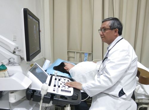
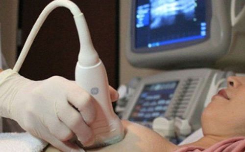
Khách hàng sử dụng dịch vụ siêu âm tại Bệnh viện Vinmec
Currently, there are many medical facilities that apply ultrasound technology in the early diagnosis and treatment of diseases. Vinmec International General Hospital system has been using the most modern generations of color ultrasound machines today in examining and treating patients. One of them is GE Healthcarecar's Logig E9 ultrasound machine with full options, HD resolution probes for clear images, accurate assessment of lesions. In addition, a team of experienced doctors and nurses will greatly assist in the diagnosis and early detection of abnormal signs of the body in order to provide timely treatment.
To register for examination and treatment at Vinmec International General Hospital, you can contact Vinmec Health System nationwide, or register for an online examination HERE
MORE:
Abdominal ultrasound is Ultrasound what parts? Things to know about echocardiography The role of ultrasound in obstetrics