This is an automatically translated article.
The article is professionally consulted by Master, Doctor Nguyen Van Phan - Department of Diagnostic Imaging and Nuclear Medicine - Vinmec Times City International General Hospital.Magnetic resonance imaging (MRI) is one of the modern and effective subclinical imaging methods, providing clear images and accurate diagnosis of the disease. Magnetic resonance imaging helps diagnose many diseases more effectively than other laboratory methods. So what is magnetic resonance imaging (MRI) and what specific meanings does it have in diagnosing the disease?
1. What is MRI?
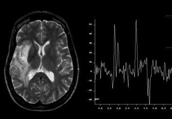
Magnetic resonance imaging (MRI) is a tomographic imaging technique that uses magnetic fields and radio waves. When the hydrogen atoms in the human body are under the influence of magnetic fields and radio waves, they absorb and release RF energy. This release is received, processed, and converted by the machine into an image.Magnetic resonance imaging with high contrast, sharp and clear, detailed, good anatomy and 3D reconstruction ability brings effective diagnosis to the doctor for the patient's pathology. In many cases, the diagnostic efficiency of MRI is much better than ultrasound, X-ray or CT scan,...
Besides, MRI does not use radiation, very safe. safety, should be highly appreciated by professional doctors in the indication of imaging and disease diagnosis.

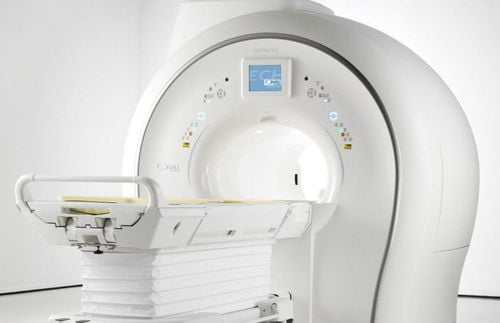
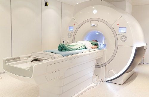
Chụp cộng hưởng từ MRI là phương pháp cận lâm sàng hiện đại.
2. Magnetic resonance imaging (MRI) is indicated for which part of the body?
Brain scan to detect brain tumors, cranial nerve tumors, cerebrovascular accident, cerebral hemorrhage, cerebral infarction, cerebrovascular malformations, traumatic brain injury, epilepsy, degenerative diseases white matter, encephalitis, meningoencephalitis, congenital malformations of the brain,...
Orbital imaging to detect lesions of the eyeball, optic nerve,...
Neck scan to detect pathological lesions such as tumors, inflammation, lymph nodes in the neck. Especially, neck magnetic resonance early detection, accurate diagnosis of damage in brachial plexus.
Spine: MRI accurately diagnoses spinal diseases, discs, ligaments such as degeneration, disc herniation, vertebral settlement fracture, disc inflammation and paravertebral software. Spinal diseases such as inflammation, spinal cord tumor, trauma,...
Abdominal - pelvic imaging to detect liver and biliary tract diseases such as liver tumors, cholangiocarcinomas, gallstones,... Diseases pancreas, spleen, kidney, adrenal gland. Diseases of the pelvic region such as colorectal cancer, prostate cancer, uterine tumors, vaginal prolapse, ovarian tumors. Especially accurately assess the stage of endometrial cancer, cervical cancer, prostate cancer,...
Musculoskeletal: MRI gives clear images of joint alveolar structures, articular cartilage , bones, tendons and ligaments. Detecting at an early stage of pathology such as inflammation, degeneration, ligament tear injury, joint effusion.
Breast MRI for early and accurate diagnosis of breast lesions such as benign, malignant and inflammatory breast tumors
MRI is one of the highly valuable methods in diagnosing fetal abnormalities, complex birth defects of the fetus. Doctors often order it in difficult cases when examining with ultrasound such as pregnant women with obesity, oligohydramnios, amniotic fluid, assessing fetal movements.
MRI is also used to diagnose heart and blood vessel diseases such as myocardial infarction, vascular stenosis, lymphatic system disease,...
3. Benefits of MRI
Patient is not affected by radiation. The patient was not biologically affected. Multi-plane image capture, easy in diagnosis. High resolution when taking soft tissue, showing better images than computed tomography (CT scan). Contrast agents have almost no side effects. It is the most effective and modern paraclinical technique available today. Fast shooting time, maximum noise reduction. Angiography without contrast injection.

Chụp cộng hưởng từ được chỉ định để chẩn đoán đa dạng các bệnh.
Vinmec International General Hospital is the first unit to put into use a 3.0 Tesla silent magnetic resonance imaging machine, bringing outstanding advantages. Magnetic resonance imaging at Vinmec will have the following outstanding advantages:
High imaging technology, first-class safety by accuracy, non-invasiveness and no radiation. High image quality, allowing doctors to comprehensively evaluate, not miss even the smallest lesions in organs. Minimizing noise, creating the most comfort for the patient when shooting, reducing stress, this helps better image quality and shortens the maximum shooting time. MRI with Silent technology is especially suitable for the elderly and children, people with weak health, patients undergoing surgery,... Can MRI at Vinmec take 3-D vascular reconstruction? requires injection of magnetic contrast, can reconstruct and handle the motion artifacts of the patient. Maximum support for specialists to perform in-depth examination, detect diseases of the skull, neck, spine, musculoskeletal, cardiovascular and cancer most accurately.
4. MRI procedure
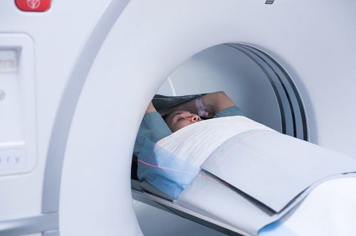
After being assigned to take an MRI from the doctor, the patient who moves to the imaging department will be greeted by the staff of the magnetic resonance room and instructed to change clothes and remove metal objects on the body. for safety during magnetic resonance imaging. When entering the imaging room, guided by the staff to lie in a comfortable position suitable for the shooting unit, the bed will automatically move to the shooting area.
Depending on the area to be photographed, the time taken for the MRI will range from 15 to 60 minutes without any discomfort. During the scan, the machine will emit all kinds of sounds, but with the higher technology MRI machine, the more noise this noise will be minimized, not causing discomfort to the photographer. The patient needs to try to lie still in a position to give the clearest and sharpest image.
In some positions and areas, the patient may be asked to hold their breath. The shooting time ends quickly without causing any discomfort or pressure on the photographer.
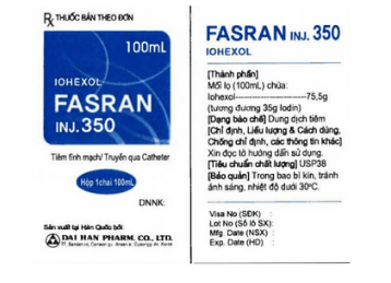
In some cases contrast injection is required from the MRI staff who will place a fine needle in the vein in the elbow area and withdraw the needle at the end of the examination.
In the case of a baby scan, the anesthesiologist will put the baby to sleep during the scan and will wake up at the end of the scan. Note that the children need to fast for ~6 hours before taking the picture, then eating and drinking normally again after taking the picture.
Please dial HOTLINE for more information or register for an appointment HERE. Download MyVinmec app to make appointments faster and to manage your bookings easily.