Most women are unaware they have a small uterus until pregnancy, when prenatal examinations and ultrasounds reveal the condition. Hearing a diagnosis of a small uterus often leads to worry, as it is commonly perceived as a uterine abnormality that might hinder pregnancy and childbirth.
1. What is a small uterus?
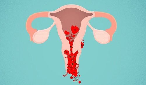
The uterus is a hollow, pear-shaped organ situated in the pelvis, between the bladder and rectum. It is divided into four parts: the fundus, the body, the isthmus, and the cervix.
The uterus begins forming at the fifth week of fetal development and grows gradually over time. It undergoes a significant growth spurt during puberty, typically around ages 13–14, when it reaches its normal physiological size. However, developmental abnormalities of the uterus may occur at various stages due to multiple factors, leading to conditions such as a double uterus, bicornuate uterus, or small uterus.
While uterine size varies among women, a standard size range is considered normal for most. A uterus is considered small if its size is smaller than the standard measurements: 3.8 cm long, 3.7 cm wide, and 2.7 cm thick. A small uterus refers to an underdeveloped organ that does not meet age-appropriate size standards.
2. Causes of a small uterus

A small uterus is an abnormality that can stem from various factors, including:
- Congenital causes: Incomplete development of the uterus during fetal growth can lead to a small uterus at birth.
- Genetics: If a mother or female siblings have a small uterus, the likelihood increases for other family members.
- Hormonal imbalances: During puberty, excessive prolactin production by the pituitary gland can impair uterine growth. Tumors in the pituitary gland may also disrupt hormonal regulation, affecting uterine size.
- Other factors: Chronic infections, toxic environmental exposures, previous ovarian removal surgeries, mumps, rubella, certain medications, excessive physical activity, or poor nutrition may also contribute to the condition.
3. Impact of a small uterus on fertility

Obstetricians classify small uterine abnormalities into three levels to assess fertility potential, predict pregnancy complications, and recommend appropriate fertility treatments:
Level 1:
- The uterus is less than 3 cm in length, with the cervix comprising about half of the total length.
- This is the most severe form, known as a rudimentary or hypoplastic uterus.
- It leads to primary amenorrhea (absence of menstruation) and infertility.
- However, if the ovaries function normally and the partner's sperm is healthy, parenthood is possible through surrogacy. Surrogacy for humanitarian purposes is legally permitted in some regions.
Level 2:
- The uterus is larger than a rudimentary uterus but maintains similar proportions between the body and cervix.
- Common signs include underdeveloped external genitalia (infantile vulva), sparse pubic hair, absence or irregularity of menstruation, severe menstrual pain, and painful intercourse.
- Hormonal treatments and metabolic interventions may help restore reproductive function in women at this level.
Level 3:
- The uterus measures 7–8 cm in length, with a 3:1 ratio between the body and cervix, consistent with normal anatomical proportions.
- This condition reflects mild uterine underdevelopment. Fertility potential and the ability to carry a pregnancy are often unaffected. However, careful monitoring during pregnancy is essential to minimize risks of miscarriage or preterm birth.
A diagnosis of a small uterus can be concerning, as it is often associated with uterine abnormalities. However, many women with this condition can still conceive and carry a pregnancy, depending on its severity. Early diagnosis and appropriate management by a healthcare professional are key to optimizing fertility and ensuring a healthy pregnancy.
To schedule an appointment at the hospital, please get in touch with the HOTLINE or book directly HERE. Download the MyVinmec App to manage, track, and schedule appointments conveniently anytime, anywhere