This is an automatically translated article.
Percutaneous kidney biopsy is the main technique used to diagnose kidney problems when other techniques cannot identify it. The results of percutaneous kidney biopsy under ultrasound guidance serve as the basis for the doctor to make a treatment plan suitable for the patient's condition.
1. Learn the technique of percutaneous kidney biopsy
Ultrasound-guided percutaneous kidney biopsy is a technique that uses an ultrasound machine to guide the collection of a sample of kidney tissue with a biopsy needle. Then, the tissue sample is examined with a microscope. Renal biopsy results suggest diagnosis and treatment modality as well as prognosis.
Percutaneous kidney biopsy is applied when other diagnostic techniques such as blood tests, urinalysis or imaging....cannot identify the disease.
2. Indications, contraindications for percutaneous renal biopsy
2.1 Indications Percutaneous kidney biopsy under ultrasound guidance is indicated in the following cases:
Hematuria, proteinuria but the cause has not been determined; Acute but rapidly progressive glomerulonephritis, or chronic glomerulonephritis with nephrotic syndrome and poor corticosteroid response; Unexplained or persistent acute renal failure (more than 1 week); Have systemic lupus erythematosus with kidney damage; Neonatal nephrotic syndrome or steroid resistance; Patients with a kidney transplant but the kidneys are not working properly, have acute or chronic rejection reactions. 2.2 Contraindications Percutaneous renal biopsy is absolutely contraindicated in patients with coagulopathy. Relative contraindications for patients with congenital kidney defects (horseshoe kidney, single kidney, ectopic kidney), polycystic kidney, abnormal renal vessels, uncontrolled hypertension pressure.

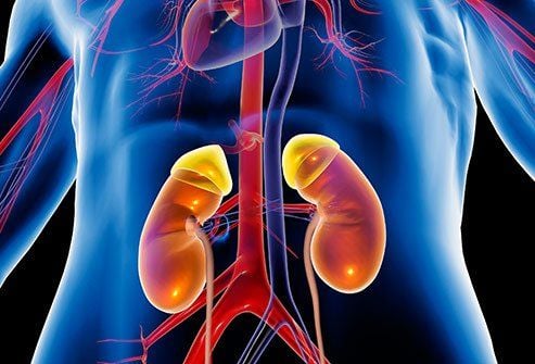
Sinh thiết thận qua da giúp bác sĩ chẩn đoán và đưa ra phác đồ điều trị bệnh phù hợp
3. Things to do before percutaneous kidney biopsy
Before performing a percutaneous kidney biopsy under ultrasound guidance, the patient should pay attention to the following do's and don'ts:
Inform the doctor about allergies, medical history, health conditions healthy, pregnant or suspected to be pregnant; Tell your doctor about any medications or herbs you are taking; You need to fast for 8 hours before the kidney biopsy. Maybe take some medicine approved by your doctor and some water; Stop using blood thinners, aspirin 3 days before the kidney biopsy; Your doctor may order blood tests before a kidney biopsy to determine if you have a clotting disorder.
4. How is a percutaneous kidney biopsy done?
Percutaneous kidney biopsy under ultrasound guidance can be performed with the following steps:
Step 1: The patient lies prone on the bed to conduct disinfection of the skin to be biopsied, then local anesthesia is administered. . Step 2: The doctor uses a knife to make a small incision on the skin, at the determined location, the biopsy needle will be inserted to take the sample. At this point, under ultrasound guidance, the doctor will precisely insert the needle into the kidney to collect a sample. Step 3: While inserting the biopsy needle, the patient is asked to hold their breath for about 20-30 seconds for the doctor to take the biopsy sample. This can be repeated until the doctor has taken enough sample to perform a kidney biopsy. Step 4: Completing the biopsy procedure, the doctor gently presses on the site of the procedure to stop the bleeding, then bandages it without stitches. The procedure takes about 1 hour. After the kidney biopsy was completed, the patient was taken to the rest room to monitor complications (if any) including: blood pressure, pulse, breathing rate, SpO2 measurement, urine color and abdominal ultrasound. In case the patient has gross hematuria, respiratory failure, blood pressure changes (increase or decrease), it is necessary to notify the doctor immediately.

Bệnh nhân được đưa về phòng nghỉ ngơi và theo dõi sau khi tiến hành siêu âm sinh thiết thận
5. Possible complications of percutaneous kidney biopsy and how to manage
Any invasive procedure is subject to complications with varying degrees of severity, so post-procedural follow-up is very important. Here are some possible complications after percutaneous renal biopsy ultrasound and how to manage:
Respiratory function is inhibited (due to sedation): Give sedation at the right dose, At the same time, carefully monitor the patient's breathing rate, SpO2 index. Use of antagonists and regimens to manage acute respiratory failure. Hypotension (due to blood loss): Monitor patient blood pressure and urine color. Treatment regimen for shock, lowering blood pressure due to blood loss. Hypertension: In case of high blood pressure, ketamine should not be used before the biopsy. Make sure your blood pressure is always within the normal range.
6. Things to keep in mind after percutaneous kidney biopsy
1 day after the procedure, the wound can be undressed and the patient is allowed to bathe normally. Do not do labor, strenuous activities, or overexertion for about 1 week after the kidney biopsy. After this time, you can eat and drink normally, but do not take aspirin. If you notice blood in your urine about 24 hours after the biopsy, you need to calm down because this is completely normal. If there is pain at the biopsy site, you should not be too worried because it is because the anesthetic has worn off, this symptom will soon go away. There may be bleeding in the kidney, when the patient needs surgical intervention to help stop the bleeding. In summary, it is very important to learn the technique of percutaneous kidney biopsy under ultrasound guidance, to help the patient prepare carefully as well as to note during and after the procedure to give accurate results. treatment facility and in good health. If you suspect that you have kidney disease, you can visit the Vinmec International General Hospital system - one of the leading prestigious addresses in the country. For the ultrasound-guided percutaneous renal biopsy technique, Vinmec uses the most modern generation of ultrasound machines today (GE Healthcare's Logig E9 ultrasound machine) with full options, high resolution transducers HD resolution for clear images, accurate assessment of lesions. In addition, the team of experienced doctors and nurses at Vinmec will help diagnose and detect abnormal signs of the patient's body early so that the most timely treatment can be given.
Please dial HOTLINE for more information or register for an appointment HERE. Download MyVinmec app to make appointments faster and to manage your bookings easily.