This is an automatically translated article.
Orbital osteosarcoma is a type of eye disease. The disease can be functional damage of the eye such as: bulging, shaking, vision loss... Therefore, orbital bone tumor surgery is considered a highly effective treatment method and prevents the ears. variables may occur.
1. What is orbital bone tumor?
The eye socket is a pyramidal cavity, the top is towards the back, the base is extended to the front, made up of the skull and facial bones. The soft tissues of the orbit do not apply directly to the periosteum but are covered by fascia. Thus, pathological processes can progress either in or out of the balance.
From the tenon to the orbital wall and the fatty tissue, there are many fibers that partially help the eyeball to stand in a certain position and easy to transport when the muscles are active, the oculomotor muscles include 4 rectus muscles and 2 muscles cross, orbital venous system, lymphatic system. This helps the eyeball to stand in a certain position in the eye socket and we can feel the pathological change from the orbit specifically here is the condition of periorbital headache.
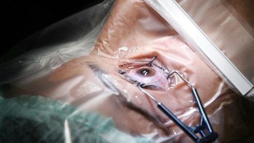
Orbital osteosarcoma is a tumor or tumor that develops from the bone wall of the eye socket. Orbital osteosarcoma surgery is performed for patients who present with symptoms caused by tumors in the eye site. In case the tumor increases in size and causes mass effect or has no clear imaging diagnosis, surgery is also indicated for orbital osteosarcoma.

Bệnh nhân sẽ được chỉ định phẫu thuật khi xuất hiện các triệu chứng do u gây nên ở hốc mắt
Before performing surgery, the patient needs to have a thorough clinical examination and imaging studies to determine the location and size of the tumor, and evaluate the structure of the base of the skull and eye sockets. To get imaging diagnosis, patients with orbital osteosarcoma are assigned to ophthalmologic examination, cranial magnetic resonance imaging, and CT scan.
Besides, the doctor will clearly explain to the patient and the patient's family about the patient's condition, the process before, during and after surgery.
However, patients with orbital osteosarcoma if their health is weak and cannot withstand the pressure of opening the skull cap will be recommended not to undergo surgery.
Like other surgeries, patients may experience some unwanted complications. Therefore, after surgery, the patient should be closely monitored.
2. Eye socket bone tumor surgery
Step 1: Preparation
To perform surgery on orbital bone tumor, the hospital needs to prepare a surgical team including: 3 doctors (1 main surgeon and 2 supporting doctors); 2 nurses; 1 anesthesiologist and 1 anesthesiologist. Full range of surgical instruments, including: endotracheal anesthesia equipment, craniotomy instruments, high-speed grinding drill with 2 mm grinding head; Microsurgery glasses, ultrasonic aspirator, neural positioning system; Consumables and closed drains placed under the skin On the patient's side, the hair on the hairline is shaved, cleaned and disinfected. Co-implementation of urinary catheter, stomach...

Đặt sonde tiểu cho bệnh nhân trước khi tiến hành phẫu thuật
Step 2: Surgery
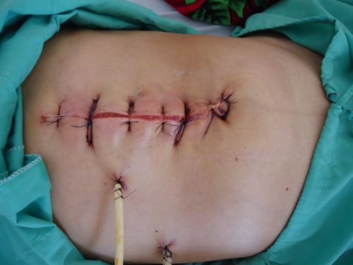
The patient is placed in supine position, head tilted 15 degrees to the opposite side. Neural navigation systems can be installed. Conduct endotracheal anesthesia with anesthetic equipment, standard fluids, blood (if necessary). The surgeon made a hairline tear from the arc to the top through the midline 1 cm. Then, dissect the skin flap, fascia and frontal fascia until the superior orbital margin is exposed, and the temporal muscle is dissected. Drill the skull, open the craniofacial lid and the ceiling of the orbit. Pay attention to the preservation of the supraorbital nerve. Depending on the position and size of the u to calculate the orbital ceiling to be cut. Pull up the sclera in the basal frontal region, exposing the orbit. Removal of osteosarcoma with chisels and drills. Hemostasis by bipolar cauterization and surgery. Replace skull cap. Step 3: Close the incision: muscle, fascia, subcutaneous, skin. End surgery
3. Post-surgery for orbital bone tumor
After surgery, the post-operative care process is extremely important. Patients should be closely monitored for pulse, blood pressure, respiration, temperature, and urine and electrolytes monitored daily. 1 week after surgery, the patient has 3rd generation antibiotics. In the case, the patient has hypopituitarism, will be prescribed corticosteroids and hormone replacement.

Bệnh nhân bị suy tuyến yên được chỉ định sử dụng corticoid để thay thế kháng sinh
Note to evaluate the possibility of retrieving the tumor, it is necessary to take resonance imaging again from 24-48 hours after surgery. Patients may experience some unwanted side effects. Therefore, the process of monitoring and handling accidents should be done as soon as possible. Complications can occur for surgery for fibromyalgia such as: Postoperative cerebral hemorrhage (epidural hematoma, intraorbital hematoma), surgical removal of hematoma if necessary; Infected surgical wound; Decreased vision; Hypopituitarism ) Vinmec International General Hospital is one of the hospitals that not only ensures professional quality with a team of leading doctors, modern equipment and technology, but also stands out for its outstanding service. comprehensive and professional medical examination, consultation and treatment; civilized, polite, safe and sterile medical examination and treatment space.
Customers can directly go to Vinmec Health system nationwide to visit or contact the hotline here for support.


