This is an automatically translated article.
Medial neuralgia is a common nerve injury. Diagnosis and early detection by means of median nerve ultrasound play an important role in decision-making and improving treatment efficiency for patients.
1. Location of median nerve
The median nerve (also called the median nerve) is a sensory and motor nerve of the arm. These median nerves extend the length of the upper limb. After arising from the lateral and medial cords of the brachial plexus, the medial nerve enters the arm in the axilla. It then travels with the brachial artery down the shaft of the humerus and into the sternocleidomastoid fossa, above the surface of the elbow joint. From the sternocleidomastoid fossa, the medial nerve crosses the forearm anteriorly and then passes through the carpal tunnel, which is a narrow passage in the palm of the wrist. After reaching the hand, the median nerve activates the various muscles of the hand.
The median nerve controls some of the major muscles of the forearm and hand, allowing for two-way communication between the brain and spinal cord, the muscles and the skin that lie above it. The brain and spinal cord can send signals through the median nerve, to the muscles it controls, with instructions about when to contract and complete specific actions. Likewise, the muscles and outer skin can transmit sensations and sensory information, such as sensations of heat and pain, through the median nerve, back to the brain and spinal cord for processing.
2. Recognize symptoms of median nerve pain
The median nerve controls many of the muscles of the forearm, providing signals to and from the brain and spinal cord. The biceps brachii is one of the muscles of the anterior forearm that is covered by the median nerve. They are involved in flexion of the forearm and wrist.
There are only two muscles of the forearm that are not controlled by the median nerve: the flexor carpi ulnaris and the flexor digitorum profundus. Instead, the flexor carpi ulnaris receives a single control signal from the ulnar nerve, and the flexor digitorum profundus receives a dual control signal from both the median nerve and the ulnar nerve. These muscles are also some of the major muscles involved in forearm and wrist flexion. In addition, a branch of the median nerve, known as the "recurrent branch of the median nerve," activates the major muscles of the hand. Thenar muscle for controlling the action of the thumb, flexion and extension. The distal branches of the median nerve innervate the sternocleidomastoid muscles of the index and middle fingers.
The median nerve is often damaged at the elbows, due to fractures of the bones of the upper arm, or wrist; due to either carpal tunnel syndrome or a wrist tear. If the median nerve is damaged in the elbow area, it is called a median nerve proximal lesion. Injury near the median nerve is often present with the blessed hand - a sign that occurs when a person is unable to execute a complete punch. It is caused by flexion of the knuckles, especially the first and third knuckles of the first and second knuckles, lost due to damage to the median nerve. Therefore, when such nerve damage is attempted, the thumb and first two fingers remain partially stretched.

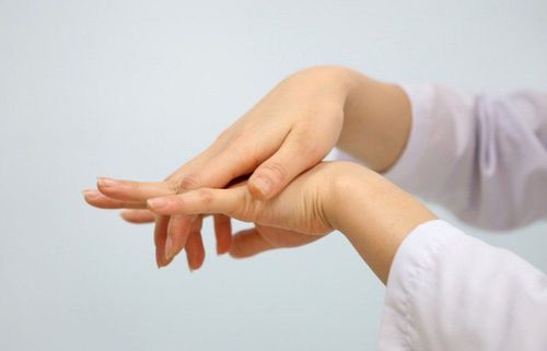
Đau dây thần kinh giữa khiến người bệnh gặp khó khăn trong cử động cổ tay
If the median nerve is damaged in the wrist, the damage is called a distal median nerve lesion. The most common cause of median nerve injury in the wrist is carpal tunnel syndrome (CTS), although a tear in the wrist can also cause median nerve injury.
Carpal tunnel syndrome occurs when a median nerve in the wrist is compressed and is often associated with pain, tingling, and numbness in the hands and arms. The median nerve is compressed due to compression between the transverse carpal and wrist ligaments. The root cause of carpal tunnel syndrome can be due to a variety of conditions, including inflammation from repeated computer use, infections, pregnancy, diabetes, and hypothyroidism.
Carpal tunnel syndrome can cause atrophy or wear and tear of the muscles in the brain stem due to lack of stimulation from the median nerve and as a result inability to function properly. Prolonged periods of time without stimulation can cause muscles to atrophy. In some cases, this can lead to the inability to move the thumb again and can cause a condition known as gibbon hand. The ape hand is characterized by severely limited thumb movement.
3. Indications for median nerve ultrasound
The median nerve at the wrist is a relatively simple structure to scan as it lies just below the transverse carpal ligament and can be found and visualized fairly easily. Nerves have a distinct ultrasonic or echogenic appearance because of their echogenic texture appearing as a combination of hypoechoic frequency bands separated by hypoechoic lines (lens) when viewed along the long axis. (vertical). In the short or transverse axis, the nerve appears mottled.
Some of the more common pathological signs of the disease will indicate an ultrasound of the median nerve, including
The size of the nerve in is more than the size of the nerve out; and cut points, such as >15mm with 2 transverse nerve regions, positive for carpal tunnel syndrome; percent change in nerve circumference in the pre-input region; Visual criteria, such as nerve deformity (folding), flattening, or hypertrophy; medial nerve mobility during wrist flexion/extension; reduced lens discrimination on short-axis views; The palmar or ventral arch is associated with nerve flattening in the distal tunnel and nerve swelling in the distal radius.

Một số trường hợp sẽ được bác sĩ chỉ định siêu âm dây thần kinh giữa
Using ultrasound to diagnose carpal tunnel syndrome (US) is an increasing trend. Advantages of median nerve ultrasound include low cost, convenience, and patient preference for this technology. Although the treatment of carpal tunnel syndrome is steroid injections, medical management, physical therapy, or even surgery on the targeted structures, median nerve ultrasound is still considered effective in determining Determine treatment options for carpal tunnel syndrome.
Vinmec International General Hospital with a system of modern facilities, medical equipment and a team of experts and doctors with many years of experience in neurological examination and treatment, patients can completely peace of mind for examination and treatment at the Hospital.
Please dial HOTLINE for more information or register for an appointment HERE. Download MyVinmec app to make appointments faster and to manage your bookings easily.


