This is an automatically translated article.
The article was professionally consulted by Specialist Doctor I Nguyen Thanh Hai - Radiologist - Department of Diagnostic Imaging and Nuclear Medicine - Vinmec Times City International General Hospital.Ankle pain can be a symptom of many conditions and diseases. Patients will suffer pain and many dangerous complications if not detected and treated promptly. X-ray of the ankle joint is one of the techniques applied to diagnose diseases of the ankle joint.
1. Why take X-ray of ankle joint
The ankle joint is one of the joints with the function of supporting the body; so the role and meaning of this part is really important to humans.Diseases related to the ankle joint cause inconvenience in moving. If not detected and treated promptly, it will leave dangerous complications later.
X-ray of the ankle joint is the most common method in the diagnosis of doctors. This technique is applied because of its quick and efficient reflection.
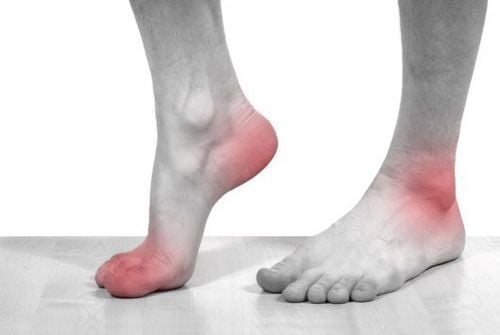
Ankle joint X-ray is indicated in case the patient has one or more of the following symptoms:
Ankle joint pain Swelling When moving the ankle joint emits abnormal noises Disease The patient experienced stiffness in the morning. Strong trauma due to impact, suspected fracture.

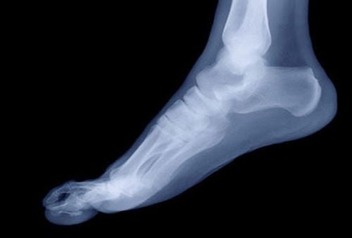
Chụp x-quang giúp phát hiện nhanh chóng các bệnh lý liên quan đến khớp cổ chân
2. Preparing for an X-ray of the ankle joint
To ensure safety and give accurate results, when performing an X-ray of the ankle joint, both medical staff as well as the patient must prepare the wood very carefully.2.1 For medical staff The person performing the ankle joint X-ray includes the specialist and the radiologist Check the dedicated X-ray machine in a ready state. Prepare and check films, film printers, storage systems, order sheets as well as results sheets for patients. Check room safety. Prepare appropriate shielding for pregnant patient 2.2 For the patient When performing an X-ray of the ankle joint or any type of X-ray, the patient should accurately report his or her condition. For example: Are you pregnant or not so that medical staff can have an appropriate treatment plan. Jewelry and metal items on the ankle joint should be removed if it affects the technique. Prepare mentally, remember the advice of doctors as well as medical staff.

Người bệnh cần tháo bỏ vật dụng kim loại quanh vùng khớp cổ chân trước khi chụp
3. X-ray procedure of straight ankle joint
3.1 Procedure for performing an X-ray of the ankle joint The following are the most common steps of the procedure for an X-ray of the ankle joint:The patient receives an order to take an X-ray of the ankle joint and moves it to the room. X-ray. The medical staff receives the order form and explains it so that the patient can understand the X-ray procedure of the ankle joint. The shooting table is adjusted so that the ball is 1m away from the film. The patient is instructed to sit on the table, the leg needs to be X-rayed, the ankle joint is extended, the heel and the back of the leg are close to the film. Leg tilt position needs to capture the outer ankle close to the film. The center ray is adjusted so that it is directed between the ankle and the film center. Pose tilt center ray above the ankle in 1cm. Stick the letter (F) (T) on the digitized sensor plate that corresponds to the leg on which the patient took the ankle joint X-ray. Medical staff enter patient information, select the corresponding program on the machine and adjust the technical factors (48kv, 20mAs).

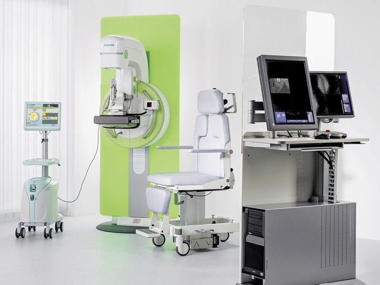
Hình ảnh máy chụp X-quang tại bệnh viện Đa khoa Quốc tế Vinmec
Any questions that need to be answered by a specialist doctor as well as customers wishing to be examined and treated at Vinmec International General Hospital, you can contact Vinmec Health System nationwide or register online HERE.
SEE MORE
Signs of bone damage on radiographs When is a routine joint radiograph performed? Sprains and ruptures of ankle ligaments


