This is an automatically translated article.
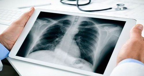
The article was professionally consulted with Master, Doctor Trinh Van Dong - Radiologist - Department of Diagnostic Imaging - Vinmec Ha Long International Hospital.An X-ray is a quick, painless test that takes pictures of structures inside the body - especially bones. X-ray beams pass through the body, and they are absorbed in different amounts depending on the density of the material they are passing through. Dense organs, such as bone and metal, appear white on X-rays. The air in the lungs appears black. Fat and muscle appear as gray.
For some types of X-ray tests, a contrast material - such as iodine or barium - is inserted into the body to provide more detail about the resulting images.
1. What are X-rays?
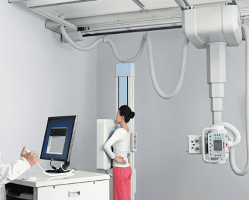
X-rays are a form of electromagnetic radiation that can penetrate solid objects, including bodies. X-rays penetrate objects more or less differently according to their density. In medicine, X-rays are used to take pictures of bones and other structures in the body. X-rays were first discovered by Wilhelm Conrad Roentgen, a German physics professor. Roentgen also studies X-rays and their ability to penetrate human tissues to create images of bone and metal that can be seen on film. To obtain an X-ray picture of a body part, the patient is positioned so that the X-rayed body part is between the X-ray source and the X-ray detector. As the X-rays pass through the body, The image appears in shades of black and white, depending on the type of tissue the X-rays pass through. For example, the calcium in your bones makes them denser, so they absorb more radiation and appear white on X-rays. Therefore, when bones are broken, the fracture line will appear as a dark area within a lighter area of bone on an X-ray. Less dense tissues such as muscle or fat absorb less and these structures appear as gray on X-rays. Air absorbs very little X-rays, so the lungs and any air-filled cavities appear black on the X-ray film. If pneumonia or tumors are present in the lungs, they are denser than the air-filled areas of the lungs and they will appear as whiter spots on an X-ray.
2. Uses of X-rays
X-rays are used in X-ray technology to examine many parts of the body, including:2.1 Bones and teeth
Fractures and infections: most fractures and infections in bones and teeth show up clearly on X-rays. Arthritis: X-rays of joints can reveal evidence of arthritis. X-rays taken over many years can help your doctor determine if your arthritis is getting worse. Cavities: Dentists use X-rays to check for cavities in your teeth. Osteoporosis: Special types of X-ray tests can measure your bone density. Bone cancer: X-rays can detect bone tumors.2.2 Chest
Lung infections or other conditions involving the lungs: Evidence of pneumonia, tuberculosis, or lung cancer may show up on a chest X-ray. Breast cancer: A mammogram is a special type of X-ray test used to examine breast tissue. Congestive heart failure: This sign of congestive heart failure shows up clearly on an X-ray. Blocked blood vessels: An injection of a contrast agent containing iodine can help highlight parts of the circulatory system that help them be seen more closely on X-rays.
2.3 Belly
Gastrointestinal problems: Barium, a contrast agent added in drinks or enema, can help detect problems in your digestive system. Swallowed item: If your child has swallowed something like a key or coin, X-rays can show the object's location.3. Risks of X-Ray Exposure
Some people worry that X-rays are not safe because radiation exposure can cause cell mutations and lead to cancer. The amount of radiation you are exposed to during an X-ray depends on the tissue or organ being examined. Sensitivity to radiation depends on your age, with children more sensitive than adults.
However, in general, radiation exposure from X-rays is low and the benefits from these tests far outweigh the risks.
However, if you are pregnant or suspect that you may be pregnant, tell your doctor before taking an X-ray. Although the risk that most X-rays pose to the unborn baby is relatively small, your doctor may consider another imaging test, such as an ultrasound.
For some people, the contrast injection can cause side effects such as:
Feeling flushed Feeling metallic taste Nausea Itching Rarely serious reactions, such as:
Low blood pressure Anaphylaxis Cardiac arrest The risk of side effects from X-rays while you're pregnant is extremely small, but it's important to always take care to protect your developing baby from harm. You should always tell your health care professional if you suspect you may be pregnant if an X-ray examination is indicated.
X-ray examination of areas of the body including arms, legs, chest, head or teeth that do not expose reproductive organs or the fetus with a direct X-ray beam. X-rays of the abdomen, stomach, kidneys, lower back, or pelvis have the potential to expose the fetus to direct X-ray exposure.

Depending on your medical condition and the area where the X-ray is needed, your doctor may cancel or postpone the X-ray examination if you are pregnant. The X-ray examination can also be modified to reduce radiation.
It is considered safe to perform X-rays while breastfeeding. Radiation does not affect milk or baby, and breastfeeding is safe after regular X-rays. Mammograms can be more difficult to see in a nursing mother, but breastfeeding women can continue to have the test when a mammogram is needed.
In the case of X-rays used with a contrast medium, breastfeeding is safe as long as no radioisotopes are used in the contrast. If radioisotopes are used, your doctor may recommend that you stop breastfeeding for a short time. You should consult your doctor about the contrast agent used and let them know if you are breast-feeding.
Radiation has some risks to consider, but it's important to remember that X-rays can help detect disease or injury at an early stage so it can be treated effectively. Sometimes performing an X-ray test can save a person's life.
The danger from X-rays comes from the radiation they produce, which can be harmful to living tissues. This risk is relatively small, but it increases with cumulative exposure. That is, the more radiation you are exposed to, the higher your risk of harm from radiation.
The risk of cancer progression is higher after exposure to X-rays. Having X-rays has also been linked to cataracts in the eyes and skin burns, but only at extreme levels of radiation. high.
Risk factors for harm to patients receiving X-rays include:
Higher number of X-ray exams Receiving X-rays at a younger age Being female (women have a higher risk than men) a bit because of radiation-related cancer) Some things you can do to reduce your risk of radiation from X-rays:
Keep track of your X-ray history and make sure your doctors know about this Ask a health care professional if there are alternatives to X-ray tests If you are pregnant or suspect that you may be pregnant, tell the radiologist or radiologist.

4. Diagnostic methods using X-rays
Many diagnostic methods use X-rays, such as: ‘
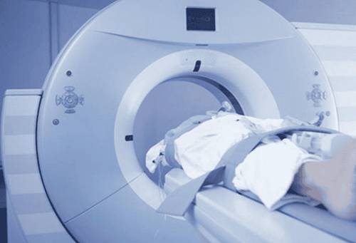
Mammography is a type of X-ray used to detect breast cancer. Computed tomography (CT) scans combine X-rays with computer processing to create detailed images (scans) of cross-sections of the body that are combined to form three-dimensional X-ray images. Fluoroscopy uses X-rays and a fluorescent screen to study moving or real-time structures in the body, such as watching the heart beat. It may also be used in combination with swallowed or injected contrast agents to view digestive processes or blood flow. Cardiac angiography uses fluoroscopy with contrast material to guide a threaded catheter inside to help open blocked arteries. Fluoroscopy is also used to precisely place instruments in certain locations in the body, such as during an epidural injection or aspiration of joint fluid. Other uses for X-rays and other types of radiation include cancer treatment. High-energy radiation at doses much higher than those used in X-rays can be used to help kill cancer cells and tumors by damaging their DNA. You do not need to prepare for a routine diagnostic X-ray. You may be asked to undress and put on a hospital gown, or at least remove any clothing on the part of your body that needs to be X-rayed.

In addition, you need to remove any metal objects on your body such as eyeglasses, jewelry or watches because they can cause interference, affecting the resulting image. If you are having an X-ray with a contrast agent such as barium or iodine, you may be given an injection with the agent before the X-ray.
If you are having a gastrointestinal X-ray, your doctor usually advises you not to eat or drink anything for 8 hours or more before the procedure so that your stomach is empty. Your doctor will let you know if you need to do this.
Vinmec International General Hospital is one of the hospitals that not only ensures professional quality with a team of leading medical doctors, modern equipment and technology, but also stands out for its examination and consultation services. comprehensive and professional medical consultation and treatment; civilized, polite, safe and sterile medical examination and treatment space.
Please dial HOTLINE for more information or register for an appointment HERE. Download MyVinmec app to make appointments faster and to manage your bookings easily.
Articles refer to sources: mayoclinic.org, medicinenet.com


