This is an automatically translated article.
The article is professionally consulted by Master, Doctor Lam Thi Kim Chi - Radiographer - Department of Diagnostic Imaging - Vinmec Danang International General Hospital. And Specialist Doctor I Nguyen Truong Duc - Doctor of Radiology - Department of Diagnostic Imaging and Nuclear Medicine - Vinmec Times City International General Hospital.Magnetic resonance imaging (MRI) is a rapid, non-invasive medical diagnostic technique that is currently being widely used in the world. MRI technology provides 3-D images, helping doctors understand information about damaged organs in the body, thereby supporting accurate disease diagnosis and timely and effective treatment.
1. What is magnetic resonance imaging?
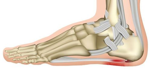
Magnetic resonance imaging, also known as MRI (short for Magnetic Resonance Imaging) is a medical diagnostic technique that creates anatomical images of the body using a magnetic field and radio waves.Magnetic resonance imaging machine is a versatile device that allows doctors to see cross-sectional images of body parts from many different angles. MRI is now commonly used in the world to examine almost every organ in the body, especially taking detailed pictures of the brain or spinal nerves.
The advantage of MRI compared to CT scanner is to clearly show the software structures in the body with high resolution, allowing clearer image recognition. At the same time, the MRI method does not use X-rays, which is safe for the patient. The information obtained from this technique is of great value to doctors in the diagnosis and treatment of diseases.
However, because the MRI patient must be placed in a strong magnetic field, patients with metal in the body such as metal fragments, bone media, etc. will cause magnetic interference and cannot be photographed. .
2. Outline of the principle of magnetic resonance

Sơ lược nguyên lý cộng hưởng từ
The particles in an atom are all in motion: neutrons and protons rotate on their own axis, electrons rotate on their own axis and revolve around the nucleus. The rotation of the aforementioned corpuscles around their axis produces an angular moment of rotation called spin. In addition, charged particles when in motion will generate a magnetic field. Because the proton has a positive charge and rotates continuously, it creates a magnetic field, called the magnetic moment. Under normal conditions, the scattered directional magnetic moments cause them to cancel each other so their signal cannot be recorded.
Thanks to these physical properties, when an object is placed in a strong magnetic field, the magnetic moments that are dissipating will change to parallel and antiparallel orientation. The object is then capable of absorbing and re-radiating electromagnetic pulses at a specific frequency. When absorbing electromagnetic pulses, inside that object will occur nuclear magnetic resonance.
Our body is composed mainly of water (accounting for 60-70%). In the composition of water molecule there are 2 hydrogen atoms (H2O). Magnetically, hydrogen is a special atom because its nucleus contains only 1 proton, so it has a large magnetic moment. That leads to a consequence: if the magnetic activity of the hydrogen atoms is used to record the water distribution of tissues in the body, it is possible to image and distinguish those tissues. Besides, in the same organ, pathological lesions will lead to a change in water distribution at the site of injury and the activity from there will change compared to healthy tissue, thereby helping to record the injury.
Applying this principle, the magnetic resonance imaging machine used a strong magnetic field and a system of radio frequency pulses to control the electromagnetic activity of the hydrogen atom's nucleus, in order to radiate energy below in the form of radio frequency signals. These signals will be captured and computer processed to create an image of the object that has just been introduced into the magnetic field.
3. Stages of Magnetic Resonance Imaging
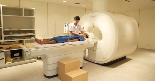
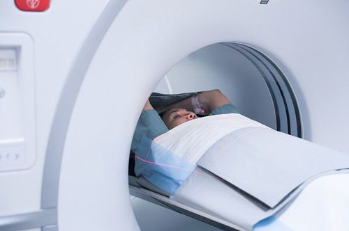
The MRI scan process consists of 4 stages:Place the patient in a strong magnetic field; Nuclear stimulation; Signal recording; Build an image from the received signal.
4. Advantages and disadvantages of magnetic resonance imaging method
4.1 Advantages Images of soft tissue structures in the body such as the liver, lungs, heart and other organs are clearer and more detailed than those produced by other diagnostic methods; MRI scans help doctors evaluate the function and structure of many organs in the body; MRI is a good tool in early diagnosis and evaluation of tumors in the body; Imaging with MRI has no side effects like routine X-ray and CT imaging; Magnetic resonance imaging allows detecting abnormalities hidden behind bone layers that are difficult to detect with other imaging methods; MRI can identify faster and more accurately than X-rays in the diagnosis of cardiovascular diseases; Does not emit radiation that is dangerous to the patient.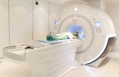
Ưu, nhược điểm của phương pháp chụp cộng hưởng từ
Please dial HOTLINE for more information or register for an appointment HERE. Download MyVinmec app to make appointments faster and to manage your bookings easily.