This is an automatically translated article.
The article was professionally consulted by Specialist Doctor I Tran Van Sang - Dermatologist - Department of Medical Examination & Internal Medicine - Vinmec Danang International General Hospital. The doctor has 18 years of experience in the field of Dermatology.Hemangiomas are the most common benign subcutaneous hemangioma in children that can be found in all regions of the body. The rate of disease occurring in infants is 59% at birth, 40% in the first month, 30% in premature babies with less than 1.8 kg of birth weight. So what causes hemangiomas under the skin?
1. What is hemangioma?
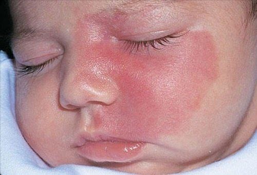
A hemangioma (scientific name is hemangioma- he-man-jee-O-muh) is a bright red birthmark that appears at birth or during the first or second week of life. It looks like a rubbery bump and is made up of extra blood vessels in the skin.A hemangioma can occur anywhere on the body, but most commonly appears on the face, scalp, chest, or back. Treatment for a baby's hemangioma (infant hemangioma) is usually not needed because it fades over time. A child with this condition in infancy usually has very little trace of growth by age 10. Treatment may be considered if the malignancy is interfering with vision, breathing or other functions.
Hemangiomas appear more often in female, white, and premature babies.
2. Features of hemangioma
Hemangiomas are characterized by endothelial cell proliferation and natural processes. Stages of hemangiomas can be divided into:Rapid proliferative phase (0-1 year): Usually lasts for 3 months, but sometimes for 6 months for superficial hemangiomas, 8-10 months for hemangiomas deep hemangioma. In this stage, 80% of hemangiomas double in size, of which about 5% grow massively, which can threaten the life, affect the function and aesthetics of the child; Stable phase (1-5 years): After the proliferative phase, the hemangioma gradually stabilizes in both size and clinical signs, lasting until 18-20 months; Regressive stage (5-10 years): In this stage, the skin color fades at first, then the subcutaneous hemangioma fades, but more slowly. This regression occurs in 70-80% of cases after the age of 6 years. The regression of subcutaneous hemangiomas is usually slower than that of cutaneous hemangiomas. There are three types of hemangiomas, including:
Capillary hemangiomas (or flat hemangiomas); Cavernous hemangioma; Mixed hemangioma. In which the flat hemangioma appears as a lipstick or a wine-colored patch on the skin due to the abnormal proliferation of the subcutaneous capillary system.
3. Causes of hemangiomas

From the embryo, due to the remnants of the embryonic mesoderm; Infection with human papillomavirus, also known as Human papilloma virus (HPV), causes uncontrolled endothelial cell proliferation of blood vessels; Due to endocrinology: High concentrations of 17-Beta Estradiol have been observed in children with hemangiomas; Heparin secreted by mast cells causes fibroblasts and endothelial cells to increase in hemangiomas.
4. Clinical symptoms of hemangioma
Hemangiomas may be present at birth, but are more common during the first few months of life. Hemangiomas in children begin as a flat red mark anywhere on the body, most often on the face, scalp, chest, or back. Usually a child has only one sign, some children may have more.During a baby's first year, the redness rapidly develops into a bump that looks like foam rubber sticking out of the skin. The hemangioma then enters a resting phase and eventually, it begins to slowly disappear.
Many hemangiomas disappear by age 5 and most will disappear completely by age 10. The skin may be slightly discolored or raised after the hemangioma disappears.
5. Is hemangioma dangerous?
The negative impact of hemangioma on humans is still being studied thoroughly. Although not life-threatening, hemangiomas can cause certain health problems.Occasionally, a hemangioma can break down and develop an ulcer. This can lead to pain, bleeding, scarring, or infection. Depending on the location of the hemangioma, it can interfere with vision, breathing, hearing, or be life-threatening - but this is very rare.
Hemangiomas can proliferate and grow continuously. In young children, if the tumor growth rate is faster than the infant's, functional and cosmetic problems such as ulcers, nasal obstruction, vision problems, and airway obstruction will obviously appear.
With adults, some special positions such as eyelids, eye sockets, parotid cause complications of compression of the optic nerve, deformed face. More dangerous can cause massive bleeding if there is trauma to the tumor area.
Although hemangiomas are benign, they also cause very serious cosmetic problems, making it difficult for patients to socialize, even autism or depression.
6. When do you need to see a doctor for hemangiomas?
Your doctor will monitor your baby's hemangioma during routine checkups. Parents should contact their doctor if the hemangioma bleeds, forms an ulcer, or becomes infected. Your child will need medical attention if the condition interferes with vision, breathing, hearing, or is life-threatening.
7. How is hemangioma diagnosed?
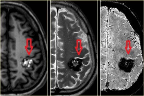
7.1 Definitive diagnosis Diagnosis is mainly based on the progression of 3 stages through follow-up, clinical examination. The most important thing is to distinguish the lesion is a hemangioma or a vascular malformation? At what stage is hemangioma? Proliferative or stable or regressive? Some diagnostic methods include:Ultrasound can help diagnose in the proliferative stage and large hemangiomas; Magnetic resonance imaging or computed tomography can be helpful in cases of hemangiomas with life-threatening complications; Angiography should be indicated when embolization is required; Biopsy: Usually not necessary because taking the patient's history and clinical findings will indicate hemangioma or vascular malformation. 7.2 Differential diagnosis of superficial hemangioma with capillary dilation or early stage of superficial hemangioma, capillary malformation: It is necessary to monitor in the first months after birth to differentiate the diagnosis. Deep hemangioma with congenital benign hemangioma: This type of hemangioma has reached peak endothelial cell proliferation at birth. This type of hemangioma is divided into two types: rapid-regressing congenital hemangioma and non-regressing congenital hemangioma. These types of hemangiomas usually do not require intervention. Benign deep hemangioma is acquired with: Angioblastoma; Kaposi-type endothelial angioma; Varicose veins; Lymphatic malformations; Arteriovenous malformations; Embryonic vascular dysplasia. Hemangiomas may not be harmful to a child's health and development, but in some cases it can interfere with a child's vision, hearing and even life. Therefore, parents still need to pay close attention to monitoring hemangiomas to promptly take their children to see a doctor if there are abnormalities.
Please dial HOTLINE for more information or register for an appointment HERE. Download MyVinmec app to make appointments faster and to manage your bookings easily.
Reference source: mayoclinic.org


