This is an automatically translated article.
The large liver fluke is a parasite that damages the liver parenchyma when people are accidentally infected. This disease also affects the whole body, causing poor appetite, weight loss and wasting. The information below will help you have more knowledge and early visit to receive proper treatment when detecting suspicious signs.1. What is the human liver fluke?
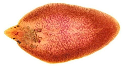
Human liver fluke is a parasitic infection, caused by two agents with scientific names, Fasciola hepattca and Fasciola gigantlca. In which, Fasciola gigantica is distributed mainly in Asia with countries such as China, Taiwan, Japan, Korea, Philippines and Vietnam. Meanwhile, Fasciola hepatica is distributed mainly in Europe, South America, Africa and only a few areas in Asia.
In the host chain to parasitize the liver fluke, human is not the main host but just a random, accidental host of the disease. Instead, herbivores such as buffaloes, cows, and sheep are the main hosts. At the same time, liver flukes also have an intermediate sign of snails living in water and mud. Therefore, when people eat undercooked snails or eat raw vegetables and leaves that grow under water such as spinach, celery, watercress, lotus root... or drink water contaminated with uncooked fluke larvae, susceptible to disease.

Các loài động vật như cừu mới là chủ ký gây bệnh
2. What are the symptoms of fascioliasis?
The clinical manifestations of the disease caused by fascioliasis are often non-specific, depending on the stage of development and the location of the parasite, as well as the number of larvae that invade the human body. Accordingly, the patient can be examined in the setting of a chronic progressive disease or hospitalized in the context of acute surgical abdominal symptoms.
The following signs are common in people with liver fluke disease:
Fatigue, weight loss, unexplained weakness. Children are stunted and malnourished. Abdominal pain in the right lower quadrant, spreading to the back or epigastrium - nasopharynx. Pain is non-specific, can be dull, sometimes severe, sometimes without abdominal pain. Poor eating, erratic, loss of appetite. Intestinal dysfunction with symptoms of nausea, vomiting, bloating, and slow digestion. There are erratic fevers such as high fever, fever with chills, or only a vague and vague fever that goes away on its own, or it can persist for an unknown reason. Chronic anemia syndrome with signs of blue skin, white nails, mucous membranes of eyes, lips, tongue pale. For cases of large liver fluke causing complications of obstruction of the secretory ducts on the gastrointestinal tract, the patient will have clinical manifestations of diseases such as obstructive jaundice, cholangitis, acute pancreatitis, gastrointestinal bleeding...
The clinical examination is the next step, the most important is to determine the direction of treatment, whether surgical intervention is needed or not. The physical symptoms on examination can be recorded as:
Painful palpation in the right lower quadrant. Determine the size of the liver is larger, the density is soft, and the pressure is painful. Sometimes there are signs of intercostal compression. Percussion has fluid in the abdomen, fluid in the pleura. If the location of the liver fluke causes an obstruction, the patient will react when pressing the abdomen with sharp pain and resistance, thinking of the possibility of peritonitis. Systemic manifestations such as parasitic infection syndrome with skin rashes, damage to other organs if the parasite is out of place...

Vàng da tắc mặt là một bệnh lý của sán lá gan lớn
3. What tests should be done in fascioliasis?
The causative agent can be detected by testing for the presence of antibodies in the serum using an ELISA test. This test does the same thing as a regular blood test. Elevated levels of antibodies to liver fluke antigens, especially IgM antibodies, suggest this diagnosis.
In addition, a stool or bile culture can help find eggs. However, the detection rate of fluke eggs is very low and depends on the testing method and the technician's experience. At the same time, care should be taken to distinguish the eggs of the liver fluke from other intestinal parasites.
In addition to the tests to determine the agent mentioned above, the patient also needs to be carried out other general laboratory tests. For a complete blood count, an elevated eosinophil count while the total white blood cell count is normal may suggest a parasitic infection.
Regarding imaging tools, ultrasound is the first-line means to examine the structures of the hepatobiliary system. Ultrasound images can show liver lesions caused by fascioliasis as honeycomb-shaped mixed echogenic foci. In addition, ultrasound also helps to identify the presence of subcapsular fluid, intra-abdominal fluid or intrapleural fluid. In some necessary cases, when it is suspected that the bile duct system is blocked for unknown reasons, a computed tomography (CT) scan of the abdomen can be performed to help investigate in more detail.

Xét nghiệm ELISA giúp chẩn đoán bệnh sán lá gan lớn
4. How to treat fascioliasis?
The group of drugs indicated for first-line specific treatment in cases of large liver fluke infections is triclabendazole, which is usually prepared in the form of 250mg tablets. Dosage is 10 mg per kilogram of body weight and only a single dose is used. Patients need to take medicine with boiled water to cool and drink after meals.
When the drug is indicated, it is necessary to pay attention to the groups of subjects with contraindications such as people suffering from other acute diseases, especially infections; pregnant or lactating women; people with a history of hypersensitivity to the drug or one of its components; people operating machinery or vehicles; Patients in the acute stage of chronic diseases of the liver, kidneys, cardiovascular... At the same time, it is necessary to inform the patient and monitor the unwanted effects of the drug that may occur immediately after taking the drug. such as:
Abdominal pain in the right lower quadrant, which can be dull or sharp. Mild fever Mild headache Nausea, vomiting Rash, itching Fortunately, in most cases, these symptoms occur only recently mild and transient, not requiring any specific treatment.
In the case of liver fluke causing damage to a mass of liver parenchyma, leading to the formation of a liver abscess, drug treatment according to the above instructions may not be effective in killing the parasites. At this time, the patient should be considered for aspiration intervention, especially for abscesses with a diameter of more than 6cm.

Nhóm thuốc triclabendazole có tác dụng hiệu quả điều trị sán lá gan
5. How to monitor patients with liver fluke after treatment?
After taking a single dose of the drug, the patient should be re-examined to re-evaluate the effectiveness of eradication of parasites. The time of follow-up is within 3 days after taking the drug and the next time is after 3 months, 6 months.
During these visits, the doctor will assess whether the clinical symptoms are reduced or eliminated, the patient's digestive ability is restored or not. At the same time, some laboratory tests were also re-assigned such as total blood analysis to evaluate the percentage of eosinophils returning to normal or decreased, liver ultrasound to assess the size of liver lesions reduced. Tests for eggs in stool or bile are absent.
In the event that a single treatment is not effective, the clinical course is unfavorable and the laboratory results are not satisfactory, a second treatment with triclabendazole should be considered. The amount of the drug is increased by 20mg/kg body weight, divided into two doses 12 to 24 hours apart. After that, the follow-up treatment is repeated as above.
It should be noted that the serum antibody test only helps diagnose the disease but does not help monitor and evaluate the effectiveness of treatment because the antibody concentration can persist for a long time after treatment. Furthermore, if imaging studies reveal large intraparenchymal lesions, abscess formation, or the site of an obstructing fluke, then an initial combination of surgical treatment should be considered. In addition, when the patient has been cured of the disease, reinfection with fascioliasis may still occur if risk factors are continued instead of actively prevented.
In summary, fascioliasis is a fairly common gastrointestinal parasitic infection in our country when the habit of eating raw is common in many regions. Early diagnosis and active treatment and prevention of reinfection are essential to restore the health of yourself and your loved ones, and avoid dangerous complications.
Reference source: vncdc.gov.vn


