This is an automatically translated article.
The article was professionally consulted by an eye doctor - Department of Medical Examination & Internal Medicine - Vinmec Hai Phong International General HospitalRetinopathy ranks second, after cataracts, in the group of diseases that cause blindness. In which, the most common are retinal detachment, diabetic retinopathy, hypertensive retinopathy, retinopathy of prematurity, central chorioretinopathy, preretinal membrane, retinoblastoma...
1. Diabetic retinopathy
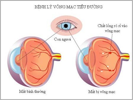
Diabetic retinopathy acquired due to complications of diabetes,. This is an eye complication that can lead to blindness. The disease will be relieved if it is diagnosed and treated in time. If you have diabetes for a long time, the higher the blood sugar, the higher the risk of diabetic retinopathy, the more severe the severity
Retinal proliferation that develops on the retinal background is the main cause of vision loss in patients with diabetes. At this stage, new blood vessels develop on the surface of the retina and optic nerve cells. These neovascularizations have the characteristic of being very fragile. When the vessel wall ruptures, blood can flow into the retina and vitreous. In the later stages of diabetic retinopathy, abnormal blood vessels and retinal scar tissue continue to grow which can cause serious complications such as retinal detachment or glaucoma.
To know if they have diabetic retinopathy or not, patients should go for diabetes screening, eye exam, ophthalmoscopy, fluorescein fluoroscopy (FFA) to support early detection of lesions of the disease. diabetic retina.
With proliferative diabetic retinopathy, the patient's vision may be blurred and the ability to perceive light may be completely lost due to hemorrhage. This disease often has no symptoms, so people with diabetes need to be screened for retinopathy as soon as they are diagnosed with diabetes and periodically visit an eye clinic for early detection and timely treatment. Avoid complicated disease progression to be treated.
2. Retinal detachment pathology

Bệnh lý bong võng mạc là tình trạng bệnh về mắt mà trong đó võng mạc bị bong ra khỏi lớp mô ở đáy mắt
Primary retinal detachment occurs due to liquefaction of vitreous fluid on the retinal surface, passing through retinal tears (appears due to retinal degeneration)
Secondary retinal detachment occurs later another eye disease (eg: diabetic retinopathy, chorioretinitis, severe myopia...).
Retinal detachment can occur in cases of trauma (closed trauma, penetrating injury, broken eyeball ...)
Retinal detachment is not painful, only disruptive visual disturbances and decreased visual acuity. Because it does not cause pain, most people are still quite subjective and do not pay attention to retinal detachment. The most recognizable symptom of retinal detachment is a gradual decrease in vision to complete loss of vision.
If you suddenly see black spots floating in front of your eyes (like flies) or see bright flashes of light, it can also be a sign of retinal detachment. Then the patient needs to go to the hospital. ophthalmologist for timely examination, consultation and treatment.
3. Hypertensive retinopathy

Một trong các biến chứng nguy hiểm của bệnh tăng huyết áp là bệnh võng mạc tăng huyết áp
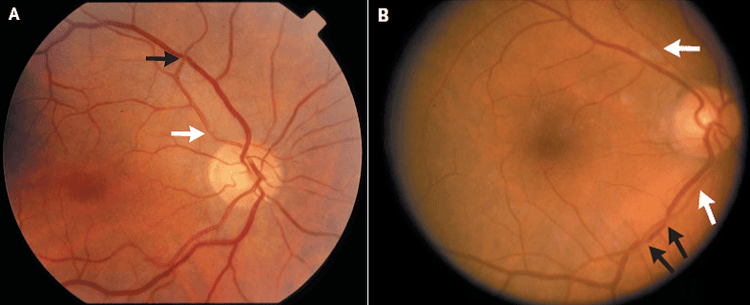
Hypertensive retinopathy progresses through many stages, which can lead to blindness if not diagnosed and treated early, with ophthalmoscopes or ophthalmoscopy doctors detect gray patches or areas white color of the retina. These areas become pale because they don't get enough blood supply. The doctor may also see bleeding from broken blood vessels or edema of the retina or optic nerve.
In people who already have hypertensive retinopathy, diabetes will make the disease worse. Controlling blood pressure is the way to limit complications. At the same time, patients with high blood pressure should have regular eye exams, which will help doctors monitor the severity of blood pressure for prevention and aggressive treatment.
4. Retinopathy of prematurity

Tỷ lệ bệnh càng cao nếu trẻ sinh càng non tháng và càng nhẹ cân
In the early stages, retinopathy in premature babies does not show up. Only in the last stage can the child's pupils become cloudy. Usually, premature and low-birth-weight infants should be examined to detect the disease at 7-9 weeks after birth. And re-examined after 3-6 months of age. Special attention should be paid to babies with birth weight of less than 3-6 months. 1000 grams or less requires re-examination every 2 weeks
In most cases, abnormal blood vessels will heal on their own (about 90%). In some children, these blood vessels are only partially healed, leading to nearsightedness or crossed eyes later in life. Sometimes, retinopathy of prematurity leaves scarring in the retina, causing vision loss. In severe cases, retinal blood vessels continue to grow abnormally and form scar tissue that causes the retina to contract, pulling it from its normal position, leading to severe vision loss.
Currently, laser retinopathy treatment has brought very positive results. The effectiveness of treatment is better if the disease is detected early and the baby has mild or moderate disease, with severe form, the treatment results are worse.
Laser purpose is to destroy abnormal retinal areas, kill new vessels and treat macular and retinal edema. However, the current common lasers still have disadvantages such as difficulty in controlling laser energy, leading to damage to areas of the retina that do not need treatment, causing extensive scarring, reduced peripheral vision, decreased sensitivity, etc. color recognition and night vision. In addition, the laser for each individual nodule makes the treatment time longer, causing a lot of pain for patients with retinopathy.
Currently at Vinmec Times City International Hospital, Pascal beam laser is being applied with many advantages. This is the most advanced retinal laser photocoagulation device in the world today, overcoming many disadvantages of today's common lasers. The laser effect time on the retinal tissues is very short, does not cause extensive burns around the laser area, so it helps to control the desired effect, causing less damage to the retina around the laser area. Besides, this is a safe method of treating the macular area. Moreover, this method causes no or very little pain, minimizing complications and side effects of the laser.
Customers can go directly to Vinmec Times City to visit or contact hotline 0243 9743 556 for support.


