This is an automatically translated article.
The article is professionally consulted by MSc, BS. Dang Manh Cuong - Radiologist - Radiology Department - Vinmec Central Park International General Hospital. The doctor has over 18 years of experience in the field of ultrasound - diagnostic imaging.Endoscopic ultrasound is an outstanding medical achievement in the field of gastrointestinal endoscopy. This is an advanced and modern technique that plays an important role in the stage diagnosis of gastrointestinal tumors.
1. What is endoscopic ultrasound?
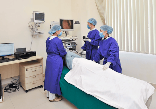
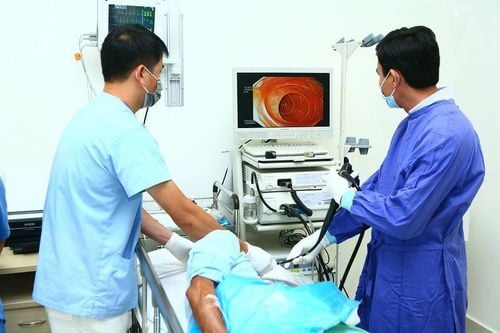
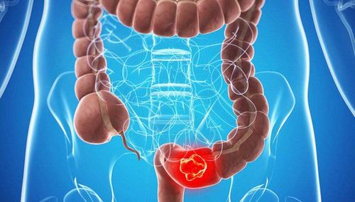
Endoscopic ultrasound is a combination of endoscopic and ultrasound techniques. This is an advanced and modern method, using an endoscope with an ultrasound probe to help diagnose and intervene in lesions of the gastrointestinal tract, bile, pancreas, mucosal and extra-mucosal lesions of the gastrointestinal tract. chemical. In particular, this technique has great significance in diagnosing cancer at an early stage (detecting tumors at an early stage helps cure up to 98-100%) or detecting tumors located deep in the colon. abdominal cavity with minimal invasiveness.Compared with other imaging methods such as multi-sequence CT or MRI, endoscopic ultrasound is more appreciated. Furthermore, the combination of endoscopic ultrasound with fine-needle aspiration biopsy allows to assess the association with neighboring nodes compared with the tumor, detect tumor invasion into surrounding organs.
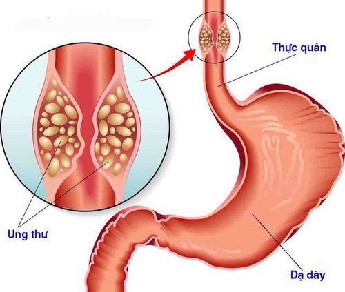
Endoscopic ultrasound has a great role and significance in diagnosing esophageal pathology, helping to evaluate the early stage for esophageal cancer, tumor invasion to the layers of the esophagus, and at the same time detect cancer. proximal lymph node metastasis, mediastinal lymph node metastasis.
For the diagnosis of gastric pathology, endoscopic ultrasonography helps in differential diagnosis of early gastric cancer, gastric submucosal tumors, and helps detect metastases in proximal and adjacent lymph nodes. compared with the tumor, detecting the invasion of the tumor into the surrounding organs, liver and spleen metastases.
Radial probe: This device gives images 360-degree image around the transducer in a plane perpendicular to the bronchoscope axis. The radial probe is used in the investigation of lesions in the gastrointestinal tract (esophagus, stomach, duodenum) and biliary tract, and lesions in the colon. Using a radial transducer is possible simultaneously for endoscopic imaging and ultrasound imaging. Linear transducer : This device captures a fan-shaped image. Under the guidance of endoscopic ultrasound combined with a linear transducer may allow fine-needle biopsies of suspected lesions. Miniprobe probe: The miniprobe probe uses a high frequency of 20 - 30 MH, which can be inserted into the lesion through biopsy of the endoscopes. Compared with radial and linear transducers, the miniprobe has the highest frequency, which helps to obtain a clear image of each layer of the gastrointestinal tract wall. Thereby helping doctors assess the early stage of cancer, effectively supporting treatment indications.
2.Indications and contraindications

3. Prepare
3.1. Person performing Specialist Doctor. Anesthesiologist Nursing. 3.2. Vehicle Endoscopic ultrasound apparatus 360° endoscopic probe and side window transducer.
4. Steps to take

5. Complications and treatment
Anesthesia complications: it is necessary to have a doctor next to you to prevent and deal with complications that occur. Perforation of hollow viscera or intra-abdominal bleeding: surgeon consultation for surgical treatment. In conclusion, endoscopic ultrasound is a great step forward in the current specialty of gastrointestinal endoscopy. This modern technique is supplementing the limitations of other subclinical diagnostic methods, thereby improving the effectiveness of more accurate and timely disease treatment.Vinmec International General Hospital is a high-quality medical facility in Vietnam with a team of highly qualified medical professionals, well-trained, domestic and foreign, and experienced.
A system of modern and advanced medical equipment, possessing many of the best machines in the world, helping to detect many difficult and dangerous diseases in a short time, supporting the diagnosis and treatment of doctors the most effective. The hospital space is designed according to 5-star hotel standards, giving patients comfort, friendliness and peace of mind.
Please dial HOTLINE for more information or register for an appointment HERE. Download MyVinmec app to make appointments faster and to manage your bookings easily.


