This is an automatically translated article.
Transthoracic echocardiography uses sound waves to create images of the heart. This method allows the doctor to see how the heart beats and pumps blood. Your doctor can use images from an echocardiogram to identify heart disease.
1. Learn about transthoracic echocardiography
Echocardiography is an important imaging tool not only in the diagnosis and monitoring of cardiovascular diseases. Echocardiography can be accessed with transthoracic or transesophageal transducers. In particular, transthoracic echocardiography is used most commonly, not only in hospitals, operating rooms, doctors' offices to portable ultrasound machines.
The basic principle of ultrasound is based on the action of sound waves. Sound waves are emitted from the transducer, passing through the chest wall, reaching the heart as well as other organs. At the same time, the transducer also receives the reflected wave, which is displayed on the computer screen. Thereby, the doctor will know what the underlying structure is like. For the purpose of describing blood flow in the heart, doctors need doppler ultrasound. This is also an ultrasound beam that is transmitted and received but will be compatible with the speed and direction of the blood flow. As a result, the doctor can detect the pathology of heart valve stenosis, abnormal flow.
2. When to do transthoracic echocardiography?
Transthoracic echocardiography is considered a routine laboratory test in middle-aged adults. Like other general tests, echocardiography should be performed once a year to assess general health status and cardiovascular function in particular. Sometimes, in the case of elderly patients, cardiovascular disease is not obvious but requires surgical intervention, echocardiography is essential to predict the tolerability of surgery as well as the postoperative course.
Particularly in Cardiology, most cases require at least one echocardiogram to diagnose pathology. Complicated cases or acute, critical ongoing disease sometimes require repeated echocardiography to assess progress, specialist consultation, or transesophageal probe access. . In clinical follow-up, echocardiography may also be indicated every 6 months to a year during follow-up visits. This is the basis for doctors to assess the patient's ability to respond to medication and adjust medication or other interventions if necessary.
Cardiovascular disease groups with indications for transthoracic echocardiography are listed as follows:
Diseases of natural valves and prosthetic valves Infective endocarditis Ischemic heart disease Acute coronary syndrome (acute myocardial infarction or unstable angina) Heart failure and cardiomyopathy Pericardial disease
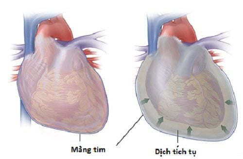
Heart tumors and mass structures in the heart Grand artery disease Congenital heart disease in children Hypertension Pulmonary disease and pulmonary vascular disease Neuropathy and other cardioembolic diseases Cardiac arrhythmias and palpitations Syncope Ultrasound disease screening Intraoperative echocardiography (including noncardiac and intracardiac surgery)
3. How is the transthoracic echocardiogram performed?
Before conducting an echocardiogram, the doctor will clearly explain to the patient and their family the goal, position, and method of implementation.
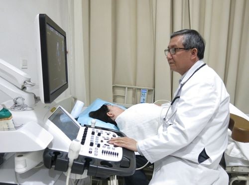
Echocardiograms can be done in a doctor's office or a hospital. The patient will lie on the bed and pull the shirt from the waist up. Your doctor will attach patches (electrodes) to your body to help detect and monitor the electrical currents of your heart.
During an echocardiogram, the doctor will reduce the light in the room so that the image can be seen more clearly on the monitor. The doctor will then apply a special gel on the patient's chest to increase the conduction of the sound waves and remove the air between the skin and the transducer (a small plastic device that sends and receives sound waves). .
The doctor will move the transducer back and forth across the chest and the sound waves that create an image of the heart are recorded on an observation screen. The patient may hear a “whoosh,” which is the sound of blood flowing in the heart recorded by the ultrasound machine.
4. What are the advantages and disadvantages of transthoracic echocardiography?
Echocardiography is the most convenient means of assessing cardiovascular function with the advantages of being quick, available, low-cost and can be performed in a specialized ultrasound room or at the bedside in the emergency case. This is also a non-invasive technique, without the risk of infection or radiation, so it is absolutely safe for pregnant women and children.
However, echocardiography is a completely subjective subclinical, depending on the qualifications, experience and skill of the performing physician. Ultrasound will also have limitations when examining patients with obesity, thick chest wall, thick subcutaneous fat, patients with chronic lung disease, patients with mechanical heart valves or distal cardiac structures. probe. At that time, the image quality is not clear and if necessary, it should be replaced with transesophageal echocardiography or other specialized imaging studies. Some patients with severe heart or lung disease and difficulty breathing may find it difficult to cooperate to perform ultrasound because the ultrasound window will be very limited.
5. Transthoracic echocardiography at Vinmec Hai Phong
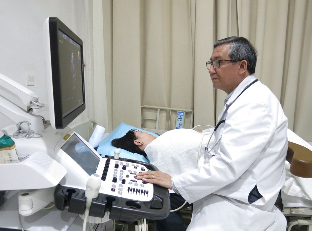
Vinmec Hai Phong International General Hospital is equipped with modern and advanced facilities and equipment and meets standards equivalent to developed countries. In particular, the echocardiogram system is a collection of completely new and high-quality machines that help echocardiography techniques be conducted quickly and conveniently.
The team of doctors who perform echocardiography at Vinmec Hai Phong have all been trained in echocardiography from basic to advanced as well as knowledge for specialized diseases, through domestic and foreign training courses. , with leading industry experts. In addition to the qualification in paraclinical imaging, the doctors also have many years of experience in clinical practice, examination and treatment of cardiovascular diseases. In addition, the nurses support patients to perform ultrasound with a dedicated and enthusiastic spirit, giving the patient a feeling of being served in the most comfortable and natural way.
The results of transthoracic echocardiography at Vinmec Hai Phong Hospital are professional and highly objective, providing accurate value for the purpose of diagnosing and monitoring cardiovascular function in particular, and the status of the heart. general health in general.
Please dial HOTLINE for more information or register for an appointment HERE. Download MyVinmec app to make appointments faster and to manage your bookings easily.


