This is an automatically translated article.
The article is professionally consulted by Master, Doctor Ta Quoc Ban - Department of Obstetrics and Gynecology - Vinmec Phu Quoc International General Hospital
Fetal ultrasound is a routine tool that helps obstetricians monitor pregnancy. Ultrasound can be performed at different times to assess the development of the baby in the womb. Along with the advancement of science and technology, ultrasound has become very popular and increasingly reflects the internal structure with 4D images. Follow the article below to learn the role of 4D ultrasound technique in pregnancy.
1. What is an ultrasound?
Ultrasound is a non-invasive, harmless test that uses ultrasound waves to evaluate the structure and nature of an organ or body (fetal ultrasound). In obstetrics, ultrasound plays an increasingly dominant role in cases where other imaging modalities, such as x-rays, are almost contraindicated.
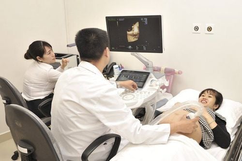
Siêu âm thai 4D
2. What is 4D ultrasound?
Today, thanks to the application of scientific achievements, ultrasound technology has made many great strides compared to the past. Compared with basic 2D ultrasound (2-plane ultrasound - 2D stands for "dimension"), 4D ultrasound allows the image displayed on the screen to be shown one more dimension in space, helping viewers Structures can be easily seen as real images, instead of through cross-sections like 2D ultrasound.In addition, the baby's structure inside the mother's uterus is not only shown visually, but also shows movements such as watching a video tape. Accordingly, the 4D ultrasound system provides more beams and the transmission speed also becomes faster. Observing the first images of the little angel, seeing clearly what the child is doing, the pregnant mother will overflow in her heart with thousands of indescribably intertwined emotions. That is one of the outstanding advantages of 4D ultrasound.
3. What is the role of 4D fetal ultrasound?
Because the nature of each type of fetal ultrasound is different, obstetricians will apply each type at each appropriate time of pregnancy, but absolutely no type shows superiority.If 2D pregnancy ultrasound only helps to evaluate the baby's development in the most basic way through the femur length, head circumference, date of birth as well as the quality and quantity of amniotic fluid remaining. ,... 4D fetal ultrasound will obviously provide more realistic images. This tool not only allows the doctor but also the pregnant mother to observe on the screen that the baby's face, limbs, and body appear through many angles. However, from a professional perspective, 4D ultrasound is a useful tool for doctors to look for abnormalities, suspected congenital abnormalities such as cleft lip, cleft palate, cleft palate, Down syndrome, etc. Short limbs or congenital heart diseases...
Not only that, coming to 4D fetal ultrasound, in addition to three-dimensional images, parents can also know what the little angel is doing in the mysterious world. his secret. Since then, some parents have kept it as the first footage of their children's life; in which, if you're lucky, you'll happen to see the baby smiling, yawning, sucking his thumb or putting his foot in preparation to kick the wall of the mother's belly.
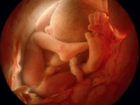
Siêu âm thai 4D cho hình ảnh sắc nét
4. When can 4D ultrasound be performed?
Depending on the purpose and stage of pregnancy, ultrasound techniques will be performed with different roles. Therefore, at the period from 20 to 24 weeks, the morphological features of the baby have developed relatively completely and this is the right time to perform 4D ultrasound.However, deciding exactly when to do it is up to the doctor who is monitoring the pregnancy. If a pregnant woman has regular antenatal check-ups, the doctor will remind the mother of the important times to assess the development of the fetus as well as survey for birth defects.
In clinical practice, doctors often appoint 4D ultrasound at 3 important times:
11-13 weeks gestation: Measure nuchal translucency and detect abnormalities early 21-22 weeks gestation : survey fetal morphology, visceral abnormalities.. Pregnancy period 31-32 weeks: assess fetal development, detect late abnormalities: duodenal obstruction, fetal growth retardation... 4D ultrasound is a useful tool to survey the fetus especially at 21-22 weeks gestation. Although ultrasound does not cause any harm to the baby, mothers should not overuse this technique because there is almost no evidence that ultrasound does not pose any risk to the baby in the future. Therefore, complying with the doctor's instructions, performing antenatal check-ups and ultrasounds at reputable places is the best measure to monitor your baby's growth day by day.
Master. Doctor Ta Quoc Ban is formerly a Lecturer in the Department of Obstetrics and Gynecology, University of Medicine and Pharmacy - Thai Nguyen University, Doctor of Obstetrics and Gynecology at Thai Nguyen Central General Hospital, Deputy Head of the Department of Obstetrics and Gynecology at the Hospital of Medical University. The doctor has full practice certificates in the field of obstetrics and gynecology such as: Ultrasound, laparoscopic surgery, endoscopic and electrocautery, artificial insemination (IUI)...
Experienced and qualified Strong in the following fields:
Obstetrics and Gynecology Ultrasound Laparoscopic surgery in gynecology Prenatal counseling Gynecological examination and consultation Infertility examination and consultation, implementation of sperm injection technique into the uterus , Pre-vaccination screening and treatment of post-vaccination reactions...
Please dial HOTLINE for more information or register for an appointment HERE. Download MyVinmec app to make appointments faster and to manage your bookings easily.