This is an automatically translated article.
The article is professionally consulted by Master, Doctor Nguyen Thanh Nam - Radiologist - Department of Diagnostic Imaging - Vinmec Danang International General Hospital.Occlusion of the lacrimal gland is a common disease in humans, occurring when the tear drainage system is partially or completely blocked. X-ray method of lacrimal gland is a highly valuable method in the diagnosis and treatment of this disease.
1. Structure of human lacrimal gland
The lacrimal gland is formed from within the skeletal system of the eyes, starting from the nasolabial fold and the eye, located between the outer nasal sprouts and the maxillary buds. Tear glands are present in the eye cavity of each person, in the upper and lower part of each eye there are tear glands. The size of the lacrimal gland is only about the size of a pea and has a flattened round shape. The lacrimal gland includes the main lacrimal gland and the minor lacrimal gland.The main lacrimal gland is located between the lacrimal area of the orbital bone wall and the eyeball. The main lacrimal gland consists of 2 parts: the cavernous lacrimal gland and a part of the lacrimal gland; The accessory lacrimal gland consists of many small lacrimal glands, located just below the human conjunctiva; In many cases when a person's eyes are irritated, they will produce a lot of tears. This tear will wash the front part of the eye, flowing down the tear duct to the sinus area. The effect of this tear makes the cornea always wet, preventing mild infections of the eye.
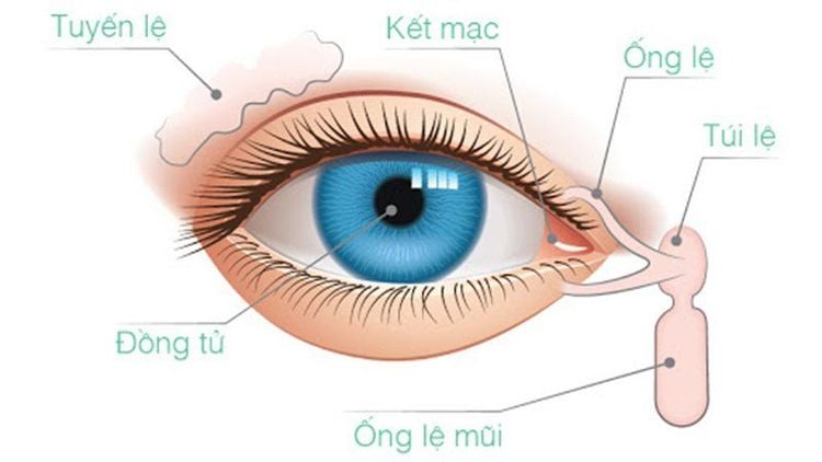
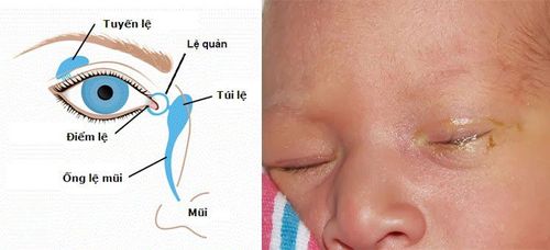
Tuyến lệ chỉ to bằng hạt đậu và có hình tròn dẹt
2. Disease of the tear gland
Obstruction of the lacrimal gland is also known as lacrimal gland obstruction. This is a common condition when the tear drainage system is partially or completely blocked, causing live tears. The disease also causes chronic eye infections.In the normal state, tears are continuously secreted from the lacrimal gland at the top of each eye. Then tears drain into two very small tear points located at the inner corners of the upper and lower eyelids and continue to flow through the two tear ducts located in the eyelids to reach the tear sacs on the side of the nose. Finally, it is led down to the nasal area through the lacrimal duct. Here tears will be evaporated or reabsorbed into the same process.
According to the results of the annual survey, about 20% of children have congenital tear duct obstruction and most of them clear up on their own after 1 year of age.
In adults, blocked tear ducts occur when the patient has eye infections, swelling, or trauma and tumors.
3. What is the effect of X-ray method of lacrimal gland?
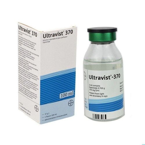
X-ray of lacrimal duct is a technique of injecting contrast material containing water-soluble iodine into the lacrimal duct to survey the ductal circulation, find the causes of ductal obstruction. For pathology of the lacrimal gland, the X-ray method is of high value for diagnosis and treatment.The lacrimal radiograph is indicated in the following cases:
The patient has frequent tearing and is suspected of ductal occlusion; Suspect stone in lacrimal gland or failed nasal routine. Contraindicated in the following cases:
Tubal inflammation Acute lacrimal gland inflammation Acute sinusitis

Chụp X quang tuyến lệ chống chỉ định với người bị mắc viêm xoang cấp
4. Preparation before performing a lacrimal mammogram
The specialist will perform a lacrimal X-ray with the help of an radiologist. Before performing the procedure, the doctor should note a few things as follows:Double check the patient's personal information such as full name, age, address,... Check the medical examination records, determine whether the patient is Do you have a history of allergies? Are you allergic to contrast or iodine-containing drugs? Explain the imaging procedure to the patient and possible complications. Patients who have a lacrimal x-ray do not need to do or prepare for the procedure.
5. Steps to take a lacrimal X-ray
The doctor adjusts the patient's supine position and puts 2 drops of anesthetic in the eye. Disinfection process must take place carefully to avoid infection later affecting the health of the patient. Dilate the lower lacrimal duct with a Bowman catheter, or use a blunt needle for the lacrimal gland. Insert the catheter in the vertical direction, after entering about 2mm, then rotate it 90 degrees to push the catheter in. When you feel a bone touch, stop. Turn the patient over to take the film. Contrast injection is carried out and then imaging is done with Waters, Caldwell, and tilt positions. During the lacrimal X-ray, the doctor must also monitor the contrast agent circulating in the duct. After 15 to 20 minutes, the contrast agent appeared in the floor of the nasal cavity and the pharynx mucosa.6. Evaluation of results after lacrimal gland scan
After performing the scan, we can observe whether the lumen of the duct is narrow or blocked, whether it is dilated on the narrow place or not; Locate the blockage in the lacrimal sac, lacrimal duct, or junction between the sac and lacrimal duct.7. Some other common problems during lacrimal X-ray
Bacterial infection: During performing a lacrimal X-ray, aseptic conditions must be observed. However, if infected, antibiotics must be used and timely treatment measures taken; In case the patient has a perforated duct, it is necessary to use a blunt needle to perform a lacrimal gland scan; Treatment of adverse events with contrast agents according to the guidelines issued by the Ministry of Health. Doctor Nguyen Thanh Nam used to be a Lecturer in Diagnostic Imaging, Hue University of Medicine and Pharmacy with good English and Russian skills and over 10 years of experience in the fields of Diagnostic Imaging.The lacrimal x-ray method is widely used today because of its high accuracy and high value in imaging results in diagnosis and treatment. To register for a lacrimal X-ray examination at Vinmec International General Hospital, you can contact Vinmec Health System nationwide, or register for an online examination HERE.
SEE ALSO:
Treating infants with blocked tear ducts Pouring breast milk into children's eyes: Do not arbitrarily Live tears due to blocked tear ducts in children


