This is an automatically translated article.
Pathology results are documents provided by pathologists. This report is intended to diagnose the disease based on pathological investigation of tissue samples taken from the patient's tumor. Based on the pathology results, detailed information about benign or malignant tumors can be obtained to help the doctor find the best treatment options.
1. What is pathology?

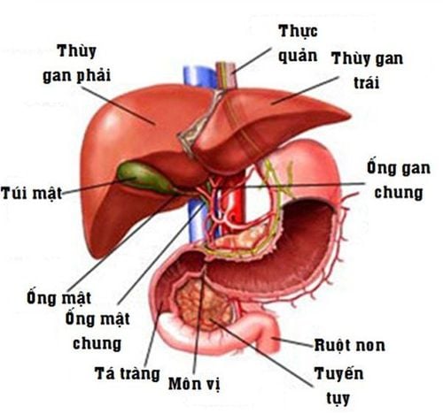
Hình ảnh giải phẫu gan,mật
Pathology is the study of tissue samples removed during surgery. They are used to diagnose diseases and decide on treatment regimens. A pathologist can provide complete consultation within the medical system, and then make an appropriate diagnosis. For example, a pathologist might study tissue removed during breast cancer surgery; This helps the surgeon decide whether to remove the lymph nodes under the arm.
Pathology includes examination of muscle tissue with the naked eye and combined with the use of a microscope. New methods in tissue and cell examination including molecular diagnostics (DNA/RNA). This involves analysis of DNA and proteins in the blood and tissues. This new technology can provide the following benefits:
Distinguish between noncancerous (benign) white blood cells and cancerous (malignant) white blood cells. Find the genetic changes that lead to cancer early. Identification of infectious agents in body tissues. Besides, along with the application of new methods, pathologists continue to use pathology atlas to see more details about anatomy, physiology, development and pathologies of the human body. , which helps the testing process and gives the most accurate final diagnosis.
2. What is the pathological result?

Kết quả giải phẫu bệnh được đưa ra để chẩn đoán bệnh dựa trên điều tra bệnh lý học về mẫu mô lấy từ khối u của bệnh nhân.
Reporting the pathology of items in a surgery is often very long and complex. It is usually divided into the following sections:
2.1 Personal information Includes patient's name, medical record number, date of completion of surgery and specimen number stored in the laboratory.
2.2 Clinical Information Includes patient information provided by the surgeon directly. It includes information about the patient's medical history and professional requests to the pathologist.
2.3 General Description It is what a researcher can see by looking, measuring, and feeling patient samples.
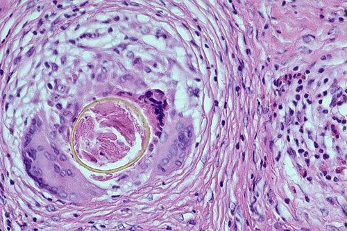
For small biopsies, this description lists things like: size, color, and density. With a biopsy or larger tissue sample such as a mastectomy for breast cancer, a longer description will be required including: size of individual tissue fibers, tumor size, how close the tumor is to margin of surgery, how many lymph nodes were found in the underarm area, and the appearance of noncancerous tissue. There must also be an exact summary of where the tissue was taken. With cytological samples, the general description is very brief, often focusing only on the number of layers or spots. If the sample is liquid, pay attention to its volume and color. 2.4 Description using a microscope The pathologist cuts tissue into thin layers, places them on slides, stains them with dye, and examines them through a microscope. The results will show the appearance of the cancer cells, how they are arranged together and how much the cancer has invaded nearby tissues in the specimen, whether they can spread into the tissue. any nearby.
To make diagnosis and treatment more effective, the pathologist compares cancer cells with normal cells. Each specific cancer will have a different scale. A tumor grade will reflect its ability to grow and spread:
First grade: Low grade or well differentiated. The cells look slightly different from normal cells. They grow slowly. Grade 2: Moderate grade or moderately differentiated. The cells are not the same as normal cells. They grow faster than usual. 3rd grade: High grade or poorly differentiated. The cells look very different from normal cells. They grow very strongly and spread very quickly. 2.5 Diagnosis This is the most important part of the pathological results, or the final diagnosis that helps the doctor to make the best treatment decision. If the diagnosis is cancer, the exact type of cancer will be noted.
2.6 Indications After the final diagnosis is made, the pathologist can provide additional information to the doctor directly treating the patient to help facilitate the treatment process and achieve high efficiency. .
The ultimate goal of pathology is not simply to describe the lesion. On the contrary, through the analysis of lesion forms, it is possible to learn the causes of the disease, explain the pathogenesis and dysfunctions caused by the lesions to contribute to the diagnosis, treatment and treatment of the disease. disease prevention.
Please dial HOTLINE for more information or register for an appointment HERE. Download MyVinmec app to make appointments faster and to manage your bookings easily.
References: cancer.org; hopkinsmedicine.org
MORE :
What is cancer? The difference between cancer cells and normal cells Is cancer diagnosis wrong? Can blood tests detect cancer?


