This is an automatically translated article.
The article is professionally consulted by Dr. Nguyen Dinh Hung - Diagnostic Imaging - Department of Diagnostic Imaging - Vinmec Hai Phong International General Hospital.
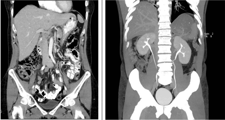
CT Scanner of the urinary system is a scanning technique that allows to observe and evaluate the entire urinary system from the calyces of the pelvis to the bladder, ureters, urinary tract, ... Computerized tomography of the urinary system Renal vasculature and urinary tract analysis can also help assess the renal arteriovenous system.
1. Overview of the urinary tract computed tomography scan to examine the renal vessels and construct the urinary tract
Computed tomography of the urinary system to examine the renal vessels and construct the excretory tract is a technique using a low-sequence computerized tomography machine to examine the entire structure and morphology of the internal organs. The urinary system includes: Kidneys, bladder, ureters, ... from the dome of the diaphragm to the pubic joint, and at the same time survey the renal artery - vein system.2. Indications for computed tomography of the urinary system to examine the renal vessels and construct the urinary tract
Computed tomography of the urinary system to examine the renal vessels and construct the urinary tract is indicated in the following cases:
● Renal colic, kidney injury.
Suspected malformation in the structure of the urinary system.
Acute or chronic ureteral obstruction of unknown etiology.
● Infections and infections of the urinary system such as: pyelonephritis, cystitis, nephritis, abscess, perirenal cavity infection, kidney parenchymal infection, ...
● Urinary tract stones: Stones bladder , kidney stones , ureteral stones .
● Diseases of the urinary system such as prostate, seminal vesicles, renal vascular disease, hydronephrosis, pelvic disease, ...
● Tumor, cancer: Kidney tumor, prostate tumor, tumor urinary system

3. Microscopic tomography of the urinary system to examine the renal vessels and construct the urinary tract
The procedure of microscopic tomography of the urinary system to examine the renal vessels and construct the urinary tract includes the following steps:
Step 1: The patient is placed in the supine position on the scanning table, arms raised above the head to avoid image noise.
● Step 2: Before injecting iodinated contrast, capture the entire urinary tract from the diaphragm to the pubic bone to locate the lesion. Assess the lesion composition including fat, or calcification, bleeding. Compare with the density of the lesion after drug injection to assess the extent of the lesion enhanced more or less. The thickness of the cut before and after injection is in the range of 3 - 5mm.
● Step 3: Inject iodinated contrast agent intravenously at a dose of 1-2ml/kg of body weight, using an electric syringe to pump at a rate of 3ml/s.
● Step 4: After 25 seconds of drug injection, a computed tomography scan of the urinary system focuses on the renal area in the arterial phase to show the renal cortex. This step allows to evaluate the vascular richness of tumor lesions, active bleeding due to renal trauma leading to drug outflow, early detection of venous drainage in case of arteriovenous malformation or vein, ...
● Step 5: After 60 seconds of drug injection, a computed tomography scan of the urinary system focuses on the kidney in the parenchymal phase to see the parenchymal phase. This step allows to assess the degree of clearance of the lesions, the enhancement of the renal vein and inferior vena cava in the case of renal tumours, and to detect lesions of the rupture or contusion in the case of trauma.

Step 6: After injecting the drug from 5-7 minutes or later depending on kidney function, computed tomography of the urinary system takes the entire urinary tract. In some cases such as ureteral stones, pyelonephritis, the doctor may perform the scan at a later time.
● Step 7: The technician re-images the urinary system, examines the renal artery and builds the excretory line by software on the scanner at the imaging chamber. The image results must clearly show the anatomical structure of the urinary system to serve the diagnosis.
All questions need to be answered by a specialist doctor as well as customers wishing to examine and perform a microscopic tomography of the urinary system to survey the renal vessels and build the urinary tract at Vinmec International General Hospital.
At Vinmec hospital, with 640 slides computer tomography machine system, it is possible to meet all imaging procedures, especially when reconstructing a complete vascular system without being affected by heart rate. The technical process is built according to international standards. KTV team is well-trained, doctors with good qualifications and experience. Patients will be welcomed, prepared and performed techniques in a fully equipped and safe hospital environment in accordance with the spirit of: PATIENT CENTRAL like the Vinmec system is building. The highest quality of diagnosis with the lowest risk is the top quality criterion that Vinmec wants to bring to patients.
Doctor Nguyen Dinh Hung has over 10 years of experience in the field of diagnostic imaging (Ultrasound, CT, MRI). Trained and practiced on hepatobiliary interventional radiology at Bach Mai Hospital (Intervention under ultrasound guidance, DSA, CT...) and deployed at the Diagnostic Imaging Department of Viet Tiep Hospital Hai Phong. Currently, he is a doctor at the Diagnostic Imaging Department of Vinmec Hai Phong International General Hospital.
Please dial HOTLINE for more information or register for an appointment HERE. Download MyVinmec app to make appointments faster and to manage your bookings easily.


