This is an automatically translated article.
The article is professionally consulted by Master, Doctor Nguyen Van Phan - Head of Interventional Imaging Unit - Department of Diagnostic Imaging and Nuclear Medicine - Vinmec Times City International General Hospital. Doctor Nguyen Van Phan is an interventional radiologist and radiologist.Ultrasound is a useful method for the diagnosis and management of benign and malignant breast disease. Ultrasound is a safe, non-invasive technique that does not use radiation. This article deals with breast abnormalities that can be detected on ultrasound.
1. Normal breast structure on ultrasound
The breast is made up of fibrous connective tissue arranged in a honeycomb-like structure that surrounds the milk ducts and the fatty tissue of the breast. The ratio of supportive buffer to glandular tissue varies widely and depends on the age, age, and hormonal status of the patient. In young women, breast tissue consists mostly of dense fibrous tissue. As you age, the dense tissue converts to adipose tissue to varying degrees, creating a mixed-fat and dense breast or an all-fat breast.The abnormal structure of the glandular tissue may appear as a tumor, because the glandular tissue is stiffer than the surrounding fatty tissue. In young women, this is a very common cause of a lump and often causes pain in the breast.
Ultrasound techniques can determine the composition of the breast: homogeneous fibrous tissue - heterogeneous tissue - or homogeneous adipose tissue.
2. Ultrasound detects abnormal lesions in the breast
The most common abnormal breast lesions usually present as a breast mass: cysts, fibroids, breast cancers, and adenomas and are palpable.2.1. Breast Cysts Cysts are the most common benign lesions in the breast, most commonly seen in women between the ages of 35 and 50. Cysts consist of fluid-filled sacs inside the breast.
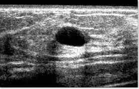
On sonography, typical features of cysts are:
Acoustic drum, clear structure Oval or round shape, thin wall Increased echo behind the cyst When fluid is stretched, the cyst becomes rounder and palpable can see. One or more cysts may be found.

Hypoechoic: Due to the cystic structure, the cyst can become rich in protein and cholesterol and contain blood and pus due to infection or hemorrhage. Thick wall: usually due to infection. Sometimes the wall can become irregular and then it can be difficult to distinguish these cysts from cancer. 2.3. Fibroblastoma is a benign tumor commonly seen in young women, especially 15-25 years of age, and rarely in women over 50 years of age.
On ultrasound, typical features of fibroadenomas are:
Clear, hypoechoic structure. A solid, oval or round, movable mass. The border is clear and sharp. The size of the transverse diameter is larger than the anteroposterior diameter. Blood flow in small vessels can sometimes be detected with color Doppler. 2.4. Breast abscess Patients often have fever, elevated white blood cells, painful accumulation of pus around the breast due to infection. The disease is mainly common in women who are breastfeeding due to inflammation.
On ultrasonography, the posterior echogenic image shows an abscessed structure containing fluid, the border may or may not be obvious.
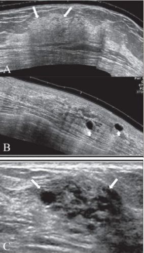
2.5. Fibrocystic change is a common benign disease in women aged 20-50 years.
The ultrasound image of the breast in this disease varies widely according to the stage and extent of the disease. The early stages of the disease may be normal, although the tumor may be palpable on physical examination. Focal fibrocystic changes may present as solid masses or thin-walled tumors. About 50% of these tumors are often difficult to identify and biopsies are required for diagnosis of malignancies.

The ultrasound image is an echogenic fat-like mass, often well-defined structure and thin capsule.
2.7. Lymphoid tumor is also known as giant fibroadenoma. This is a rare lesion, common in women over 40 years old. Most are benign, only a small percentage can turn into malignant tumors.
Image of phyllodes tumor on ultrasound:
The sound structure is often heterogeneous, with hypoechoic. Round or oval in shape, in the case of malignant lesions the margins may be irregular. Back wall lightening, no internal calcification. 2.8. Breast cancer Breast cancer is the most common malignancy in women. A woman's risk of breast cancer increases with age. Most women diagnosed with breast cancer are over the age of 50, but younger women can also get breast cancer.
Common clinical symptoms of breast cancer are:
Tumor or thickened breast tissue. Nipple dimpling or skin indentation around the breast. On ultrasound, the typical features of breast cancer are:
There are hypoechoic masses of irregular shape, sometimes round or oval, and oriented not parallel to the skin. Indistinct boundaries are sometimes bounded. Possibly posterior dorsal shadow and minor calcifications. The above are common lesions in the breast on ultrasound. Although it is not possible to distinguish all benign and malignant lesions in the breast on ultrasound, ultrasound plays an important role in the detection of some low-risk and malignancies. monitor tumor growth without the need for more invasive procedures such as biopsies.
Vinmec International General Hospital offers customers a Breast Cancer Screening Package to help check and screen for signs of breast cysts as well as the risks of breast cancer. The package includes diagnostic methods of bilateral breast ultrasound and mammography for accurate results, assisting doctors in the examination.
Please dial HOTLINE for more information or register for an appointment HERE. Download MyVinmec app to make appointments faster and to manage your bookings easily.


