This is an automatically translated article.
The article is written by Master, Doctor Mai Vien Phuong - Gastroenterologist - Department of Medical Examination & Internal Medicine - Vinmec Central Park International General Hospital.Invite you to follow the series on treatment of diaphragmatic hiatus hernia by Doctor Mai Vien Phuong:
Treatment of diaphragmatic hiatus hernia part 1: Medical and surgical treatment Treatment of hiatal hernia part 2: HIS angle shaping method Treatment of diaphragmatic hiatus hernia part 3: Robotic surgery and laparoscopic surgery "HIS angle" refers to a normal acute angle formed between the abdominal esophagus and the fundus of the stomach (large bulge or fundus) at the gastroesophageal junction (esophagogastric junction). This angle is one of the important factors in preventing gastroesophageal reflux disease. When the fundus is enlarged by air or other gastric contents, the thoracic esophageal structures are "pushed" from left to right, closing the gastroesophageal flap valve.
1. HIS . angle forming methods
1.1 Shaping the HIS angle according to NISSEN The bulge is wrapped around the oesophagus 360o.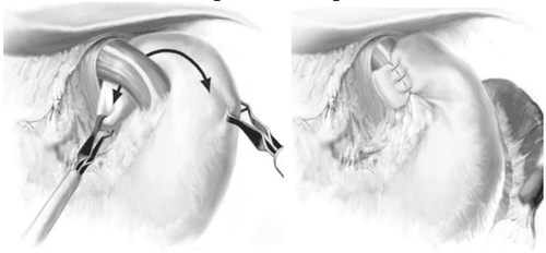
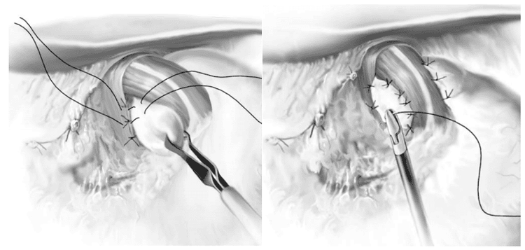
Primary esophageal motility disorders: scleroderma, achalasia. Esophageal motility disorders secondary: decreased esophageal motility leading to Barrett's esophagus or chronic reflux. Inability to tolerate complete His angle reconstruction: dysphagia, flatulence, chronic nausea, modification of an obstructing 3600 image, dysphagia. 1.2 Shaping the HIS angle according to TOUPET The aneurysm is incompletely sutured (270o).

2. The role of His angle shaping
His angle shaping to prevent reflux or not is still under discussion. The authors do not support his angle reconstruction because of the significant complication rate after surgery, as well as the longer duration, increased surgical costs. However, most authors advocate routine His angle reconstruction after repair of the diaphragm defect. There are many reasons for this. The 24-hour esophageal pH test showed increased esophageal exposure to gastric juice in 60-70% of patients for sliding diaphragmatic hernia, and 71% for paraesophageal hernias. Furthermore, there was no relationship between the patient's symptoms and cardiac tone. Finally, esophageal dissection can lead to postoperative reflux even though the patient did not have reflux before surgery.3. How to shape His angle?
There are two ways to shape His angle, which is to create a full His angle and to create a partial His angle.Partial His angle shaping technique is indicated for cases such as scleroderma, achalasia, reduced esophageal motility leading to Barrett's esophagus or chronic reflux, inability to tolerate with complete His angle reconstruction: dysphagia, flatulence, chronic nausea, modification of an obstructive 3600, dysphagia.
One of the most commonly applied methods of suturing the gastric aneurysm is the Nissen surgery. Nissen surgery has a low complication rate and shorter hospital stay compared to open surgery. However, the rate of patients with swallowing choking and gas in the stomach after surgery is quite high. To reduce the risk of choking and gastric distention, DeMeester and Peters suggest placing a bougie in the esophagus (endoscopically) during surgery, while reducing the length of the coil and the mobility of the stomach. more than.
Toupet surgery is a modification of Nissen, in which the gastric aneurysm is sutured incompletely (270 degrees), with the aim of reducing the incidence of postoperative cardiac obstruction.
In fact, among the methods of partial His angioplasty, the choice of method is up to the expertise of the surgeon. Our main His angle shaping technique is the Dor method because this is an easy method to perform, giving a lower rate of postoperative choking complications compared to other methods.
4. Place the puzzle piece
There are many methods of placing puzzle pieces. According to Oelschlager BK., a U-shaped piece about 7 x 10 cm was placed in the diaphragm after stitching the two diaphragms. The graft is fixed to the diaphragm with bio-glue or removable sutures.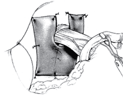
Please dial HOTLINE for more information or register for an appointment HERE. Download MyVinmec app to make appointments faster and to manage your bookings easily.


