This is an automatically translated article.
Article by Master, Doctor Mai Vien Phuong - Gastrointestinal endoscopist - Department of Medical Examination & Internal Medicine - Vinmec Central Park International General Hospital.
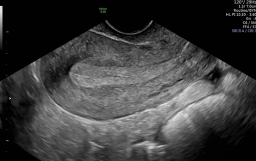
Uterine fibroids are a common gynecological disease of all ages, especially women in the period of childbearing, pregnancy or menopause. The location of fibroids can be subserosal, in the lining of the uterus, in the muscle layer of the uterus, or outside the uterus. Ultrasound is a safe, non-invasive imaging technique widely used in the diagnosis of uterine fibroids.
1. How is an ultrasound of the uterus done?
There are 3 types of ultrasound that can diagnose uterine fibroids, which are abdominal ultrasound, transvaginal ultrasound and uterine pump ultrasound.
Transabdominal ultrasound:
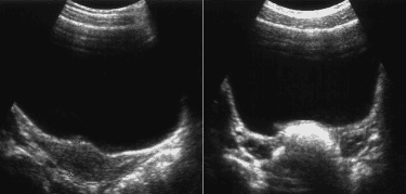
In most cases, the patient will lie supine on the examination table. The doctor may ask the patient to tilt left or right in some cases for better examination. After the patient lies on the bed, the doctor will apply a gel to the abdomen and pelvis to help the ultrasound waves pass through the body easily, thereby recording images of the pelvic organs. You may feel some discomfort as the ultrasound transducer presses against your abdomen and pelvis. After the exam, you will wipe your abdomen with a clean towel or paper. Transvaginal transvaginal ultrasound:
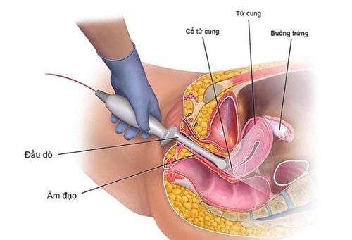
is a widely used technique in gynecological ultrasound. The patient will urinate cleanly before performing the ultrasound. Then, the patient is instructed to lie on the examination table in the same position as the gynecological examination position. The doctor will insert a small specialized probe into the patient's vagina to take pictures of the pelvic organs. Uterine pump ultrasound:
Similar to transvaginal ultrasound, the only difference is that the patient will be pumped a little physiological saline into the uterus through the cervix, then perform an ultrasound.

2. The benefits and limitations of ultrasound in the diagnosis of uterine fibroids
The benefits of ultrasound in the diagnosis of uterine fibroids:
Ultrasound is a non-invasive imaging technique (no needles or injections are used). The implementation time is faster than other uterine fibroid screening techniques such as computed tomography, magnetic resonance, hysterosalpingogram... With ultrasound, the doctor can perform a multitude of things. Different cross-sections to evaluate u comprehensively. Occasionally, an ultrasound exam can cause temporary discomfort, but no pain. Ultrasound is widely used and less expensive than most other imaging methods. Ultrasound scans give clear pictures of the uterus and ovaries. From there, it helps to diagnose uterine fibroids in any position, size, structure, and perfusion properties. Differential diagnosis of uterine fibroids and some other pelvic pathologies such as ovarian cysts eggs, diseases related to the bladder, rectum... Ultrasound images are extremely safe and do not use radiation. Therefore, this technique can be used repeatedly to monitor the progression of uterine fibroids. Limitations of ultrasound in diagnosing uterine fibroids:
In some cases, abdominal ultrasound is difficult to evaluate the uterus and ovaries due to many stools and air in the colonic lumen. In this case, if the patient is married or sexually active, the doctor may choose to use transvaginal ultrasound for evaluation. Sometimes both abdominal and vaginal ultrasound cannot confirm the diagnosis of uterine fibroids or accurately assess the location, structure, and nature of the tumor as well as the effect of the tumor on the organs. If the diagnosis is contiguous, or the differential diagnosis is required from other conditions, the alternative imaging modality, magnetic resonance, is often indicated for further evaluation. In summary, uterine fibroids are a common gynecological disease of all ages, especially women in the reproductive period, pregnancy or menopause. Uterine ultrasound is a safe, non-invasive imaging technique widely used in the diagnosis of uterine fibroids.
In order to help customers detect and treat other gynecological diseases early, Vinmec International Hospital has a basic gynecological examination and screening package, helping customers detect early inflammatory diseases Easy, inexpensive treatment. Screening detects gynecological cancer (cervical cancer) early even when there are no symptoms.
Basic gynecological examination and screening package for female customers, has no age limit and may have the following symptoms:
Abnormal vaginal bleeding Having menstrual problems: irregular menstrual cycle, irregular menstrual cycle Irregular vaginal discharge (smell, different color) Vaginal pain and itching Female clients have several risk factors such as poor personal hygiene, Unsafe sex, abortion,... Female customers have other symptoms such as: Abnormal vaginal discharge, itching, pain in the intimate area, abnormal vaginal bleeding. When registering for the Basic Gynecological Examination and Screening Package, customers will receive:
Gynecological Specialist Examination Transvaginal Uterine Ovarian Ultrasound Vaginal Bilateral Breast Ultrasound Tests such as: Treponema pallidum test rapid, Chlamydia rapid test, taking samples for cervical-vaginal cytology, bacterioscopic staining (female vaginal fluid), HPV genotype PCR automated system, Total urinalysis by automatic machine.
Please dial HOTLINE for more information or register for an appointment HERE. Download MyVinmec app to make appointments faster and to manage your bookings easily.


