This is an automatically translated article.
This article is written by Dr. Nguyen Thu Huong - Deputy Head of Diagnostic Imaging - Breast Cancer and Benign Disease Screening Unit - Vinmec Times City International Hospital.
There are many imaging modalities used to detect and diagnose benign breast pathologies and also breast cancer, mammography or mammography is a proven screening method for breast cancer. reduce mortality from breast cancer. There are also a number of other imaging diagnostic tools such as ultrasound, magnetic resonance... which coordinate both in screening and diagnosis.
1. What is a mammogram?
Mammography is a method of using low-energy X-rays to image the structure of the breast, helping to find breast cancer. If you are advised to use a mammogram and are preparing to have a mammogram, the following knowledge is essential.
It is necessary to have a mammogram to detect breast cancer at an early stage, even if the mass is not palpable, so that it can be treated in the most timely manner.
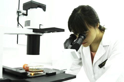
Chụp X-quang vú giúp chẩn đoán ung thư sớm
2. What is the purpose of using a mammogram?
Mammograms are used in screening and diagnosis. Screening is used in large numbers of people when there are no symptoms with the aim of detecting breast cancer as early as possible. For the purpose of disease diagnosis, when the patient has symptoms detected in the breast, they will be examined and ordered to take a mammogram or when abnormality is detected on the screening film, mammogram of additional positions. will be indicated to see the lesion more clearly.

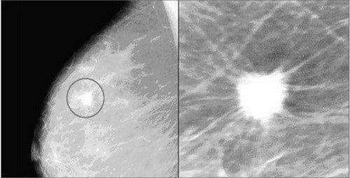
X-quang vú cho phép chẩn đoán hình ảnh về ung thư vú
3. Symptoms can be found on mammograms
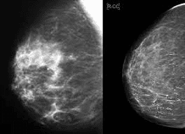
Breast X-ray is used to detect breast abnormalities, this is not a gold standard tool in diagnosing cancer, but based on this tool, doctors can detect suspicious sites. and specify the necessary interventions to take tissue samples for testing if necessary. Two typical symptoms commonly seen on mammograms are calcifications and masses.
Calcification : Is calcium deposits in the breast parenchyma with small size, bright spots on X-ray film, there are two types: large gross calcification and microcalcification. Large gross calcifications, usually about 1 mm or more in size, are caused by the deposition of calcium crystals in the blood vessels of the breast, due to old trauma or inflammation. Usually gross large calcifications are benign in nature and do not require biopsies, and are often present in elderly women. Microcalcifications are small calcifications < 0.5 mm located in the mammary gland parenchyma. When present, microcalcification should be noted because it can be a symptom of breast cancer, but not 100% breast cancer. If the morphology and distribution are in doubt, the doctor will order a biopsy to diagnose the pathology.
Mass: A mass is an area that appears to have increased optical density on a mammogram, with a different shape and border compared to the breast parenchyma. With or without microcalcification, the contour and shape of the mass should be carefully evaluated for signs of cancer. Masses seen on mammograms may be cysts or solid masses. Often, combined with ultrasound will help doctors have more information to diagnose a mass detected on a mammogram.
Vôi hóa tuyến vú là một trong những hình ảnh được tìm thấy trên X -quang vú
4. How is the mammogram?
The photographer will be dressed and taken by a specialized machine that emits X-rays with lower energy than conventional X-rays. The patient's breast will be gently pressed to thin out the parenchyma with a compression table with X-ray generator and a lower support table with video recording. For routine screening and diagnostic purposes, the patient will be photographed in both upper-lower and lateral-internal positions in both breasts for comparison. For further diagnosis, the doctor will prescribe additional positions such as focal compression and magnification ...
Preparation before mammogram:
If you have a choice, go to a facility that can Fully equipped with standard equipment, well-trained doctors and experts in diagnosing mammograms. Stay at the same facility that guarantees repeat stays each year, so it's easier to compare your before and after results, If you're going to be screened at another facility, bring enough Papers and results from previous visits, especially if there are pathologic results after the biopsy. It is advisable to schedule mammograms when your breast parenchyma is not engorged and tender to reduce discomfort and obtain good quality films, trying to avoid the week immediately before your period. On the day of the shoot, do not use deodorant underarm roll because it affects the quality of the film, after taking the photo you can use it. You should wear loose pants and shirt to just take off the top, avoid wearing a skirt. Talk to your doctor if you notice any abnormalities in your breasts before the scan. What should be informed to the technician when taking a mammogram?
Always tell them if there is any change in your mammary gland, you can also remind the imaging technician if you have had surgery before, there are scars... Before the scan, it is important to inform if you have pain, you have breast augmentation, you are breastfeeding or could be pregnant...

Phụ nữ có thai cần thông báo với bác sĩ trước khi chụp X-quang
5. Are mammograms safe?
A mammogram is a method of using X-rays, but the benefits of a mammogram far outweigh the concerns about X-ray absorption. Modern machines today use X-rays with low energy but with good image quality. On average, a single radiograph has a low dose of about 0.4 mSv, much smaller than a chest X-ray. For a better understanding, it is common for a person to absorb about 3 mSv a year in the background radiation. So the radiation dose for a mammogram is equivalent to a woman absorbing normal radiation in the environment for about 7 weeks.
If you become pregnant or suspect that you may be pregnant, tell your doctor or the imaging technician right away. Although the risk to the fetus is very small, mammograms should be considered in this case. Mammograms are not used for screening purposes in pregnant women.

Chụp X -quang vú có nhiều ưu điểm và được áp dụng phổ biến
6. Understanding when receiving mammogram results
The breast radiologist will read the film and answer the results for you, usually the patient will know his previous classification results and his mammary parenchymal density and inform the doctor in advance . So, what is the resulting classification?
BIRADS : This is a classification system of results that tells you if you have any imaging symptoms after screening and what you need to do next:
BIRADS 0: Visit not finished, need to do more tests Additional tests such as additional orthopedic scans or in combination with ultrasound or injections BIRADS 1: Negative, you return for routine screening annually or as indicated by your physician for follow-up screening BIRADS 2: Damage benign lesions, you return for routine yearly screening or as indicated by your physician for follow-up screening BIRADS 3: Lesions are more likely to be benign (> 98% are benign), you need to be monitored periodically, once every 6 months for two years to evaluate the size and nature of the BIRADS 4 lesion: Suspected malignancy (2-95% malignancy), you need to be biopsied to implementing the quadrants. BIRADS 5: Lesions with high suspicion of malignancy (>95% malignancy), you definitely need an early biopsy for a definitive diagnosis. BIRADS 6: Malignant lesions have been biopsied with known results, often this case for diagnosis and evaluation after conservative treatment. Mammography is a safe, affordable method with sufficient scientific basis to be used in early breast cancer diagnosis, effective treatment and cure. Choosing a secure mammogram is very important to help you get good results. Understanding the results of mammograms will help you know and plan for regular checkups, good and reasonable health care.
In January 2020, Vinmec Health System established a Cancer and Benign Breast Cancer Screening Unit to turn Vinmec Times City General Hospital into a Center for screening, diagnosis, treatment, and consulting on cancer. The leading modern and comprehensive breast cancer and benign breast diseases in Vietnam.
The Breast Cancer and Benign Disease Screening Unit is the place to deploy a uniform and standard breast cancer screening model with a team of specialized doctors and a modern machine system that fully meets the needs of patients. international standards in screening.
Counseling on breast cancer, benign breast diseases, screening methods, clinical examination and instructions for breast self-examination for clients; Screening, diagnosing breast cancer and benign breast diseases through the means of mammogram X-ray, breast ultrasound, breast MRI, PET/CT; Deploying interventional techniques under the guidance of images such as: cell puncture, biopsy, vacuum aspiration, cyst destruction, locating the wire needle under the guidance of X-ray, ultrasound; Participate in multi-specialty consultations, coordinate with oncology centers in monitoring after breast cancer treatment. To book an appointment, you can go directly to the Breast Cancer and Benign Disease Screening Unit at Vinmec Times City International Hospital or contact the Breast Clinic's hotline: 0910.704.068.
Please dial HOTLINE for more information or register for an appointment HERE. Download MyVinmec app to make appointments faster and to manage your bookings easily.